How To Read Bladder Ultrasound
Ultrasound of the bladder is appointed as identify various pathologies residual urine volume and to control the flow of the treatment process. Various body tissues conduct sound differently.
Ultrasound Estimated Post Void Residual Bladder Youtube
An empty bladder is required.

How to read bladder ultrasound. However a person may need to hold their urine so that the bladder is full for part. The bladder ultrasound is painless. Whether you are pregnant or not the rules are the same for a transvaginal ultrasound exam.
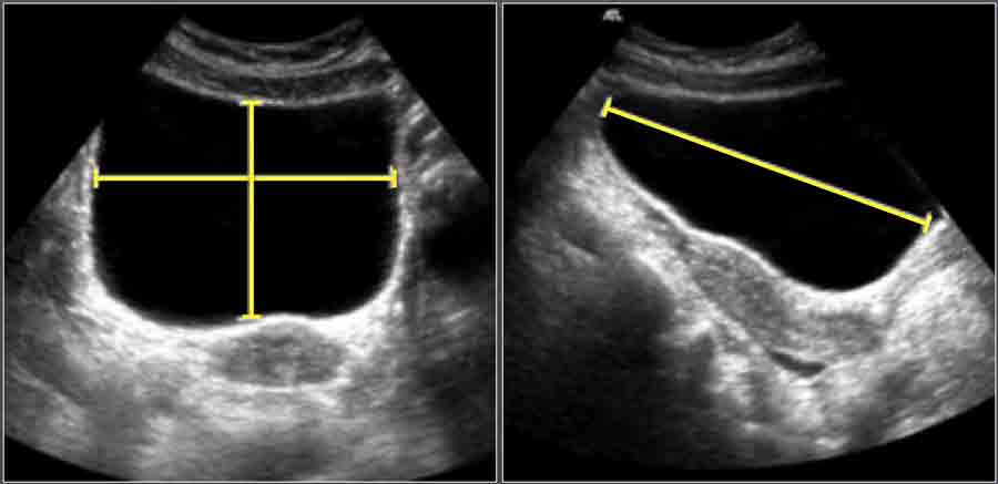
To evaluate for ureteral jets properly make sure you are in the transverse view of the bladder and decrease your color Doppler scale to about 10-20cms or you can use power Doppler. Both transverse and longitudinal images are typically assessed. C When you find the ideal ultrasound bladder image press the probe button again.
As an alternative to bladder catheterisation it has been shown to significantly reduce urinary tract infection UTI as well as increasing patient comfort and satisfaction. The advantages of ultrasonography of organs of the urogenital system are the simplicity high. Transvaginal ultrasound requires covering the ultrasound transducer in a plastic or latex sheath which may cause a reaction in patients with a latex allergy.
A kidney and bladder ultrasound or renal ultrasound uses high frequency sound waves transmitted through a transducer probe to visualize and assess your kidneys ureters small muscular tubes that join the kidneys with the bladder and urinary bladder. The transducer uses sound waves to make images of your bladder. During the examination an ultrasound machine sends sound waves into the kidney area and images are recorded on a computer.
Rock the probe so that it points down towards the pelvic cavity. The ureters are thin tubes that carry the urine from the kidneys to the bladder. Press the Scan button a Press the probe button to start ultrasound scanning to locate the bladder b Make sure the ultrasound bladder image is the biggest and centered.
The TV provides a more magnified view so a full bladder just gets in the way. Your child may feel a slight pressure as the transducer is moved over the abdomen. You will lie on a table.
A beep is heard once the calculation is done. Locate the bladder and get the most central and biggest image you can get on the ultrasound scanner screen. To read an ultrasound picture look for white spots on the image to see solid tissues like bones and dark spots on the image to see fluid-filled tissues like the amniotic.
The test is not painful. It will not penetrate bone like an X-Ray. Ultrasound is a non-invasive immediate tool used to image tissue.
What will happen during a bladder ultrasound. During a transabdominal ultrasound you may experience discomfort from having a full bladder or lying on the examination table. Ad Symptoms include blood in the urine frequency and urgency of urination.
Ask your child to lie still during the procedure so the sound waves can produce the proper images. The ultrasound probe is positioned in the suprapubic region pointing towards the bladder. Inside the vagina the probe is closer to your organs allowing us to see them more clearly.
Methodology of ultrasonic studies of the bladder refers to the most common effective and efficient methods of diagnosis. Then he will take more images of your bladder when it is empty. Ultrasound Ureteral Jets are a color Doppler finding during bladder ultrasound that detects the flow of urine into the bladder at the level of the trigone.
He will ask you to urinate. Some tissues absorb sound waves while others reflect them. Portable bladder ultrasound scanning technology offers a non-invasive fast and painless method to measure urine volume in the bladder.
The 10 Most Important Warning Signs and Symptoms of Bladder Cancer You Should NOT Ignore. This video educates about how to see the gall bladder gall bladder calculus gallbladder wall oedema and common bile duct. He will then move a device called a transducer over that area.
Your healthcare provider will put gel on your lower abdomen. The black-and-white images show the internal structure of the kidneys and related organs. The technician may ask your child to lie in different positions or hold his or her breath briefly.
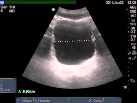
The ultrasonographer inspects for free fluid outside of the bladder. Bladder Ultrasound Longitudinal View Place the transducer with the indicator pointing towards the patients head in the patients midline right above the. When you have this image press the probe button to calculate urine volume.
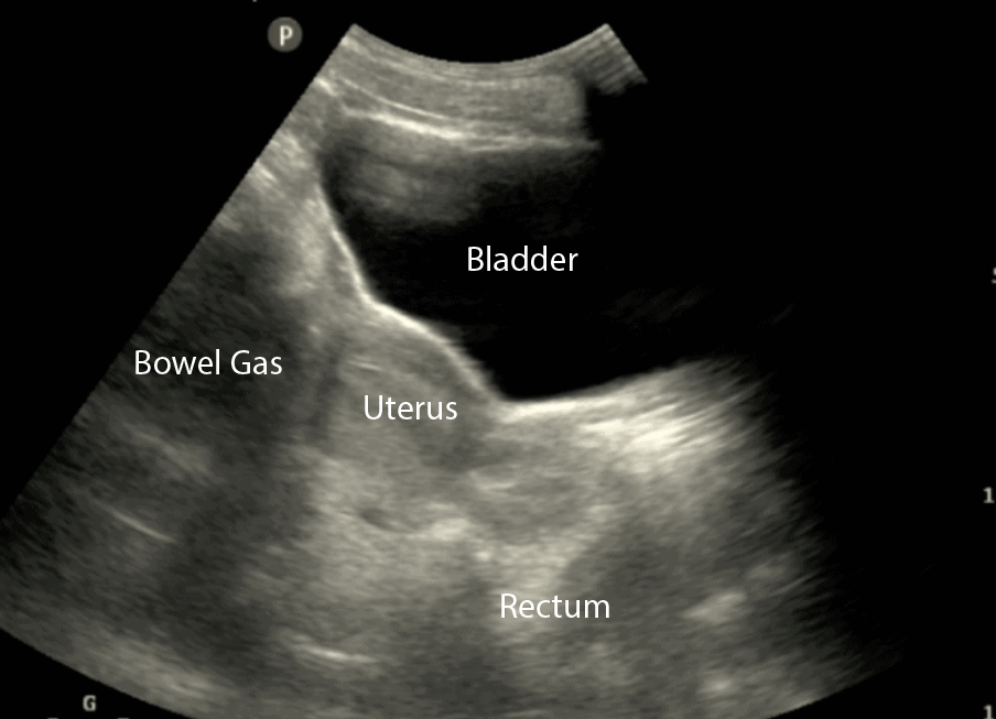
A bladder ultrasound is a test that uses sound waves to produce images of the bladder. So the first step to help you read the ultrasound image is to be familiar with the anatomy that you are imaging. Pad Scan Bladder Scanner will start calculation automatically.

Radiology Quiz Urology News

Small Animal Abdominal Ultrasonography The Urinary Tract Urinary Bladder Urethra Today S Veterinary Practice
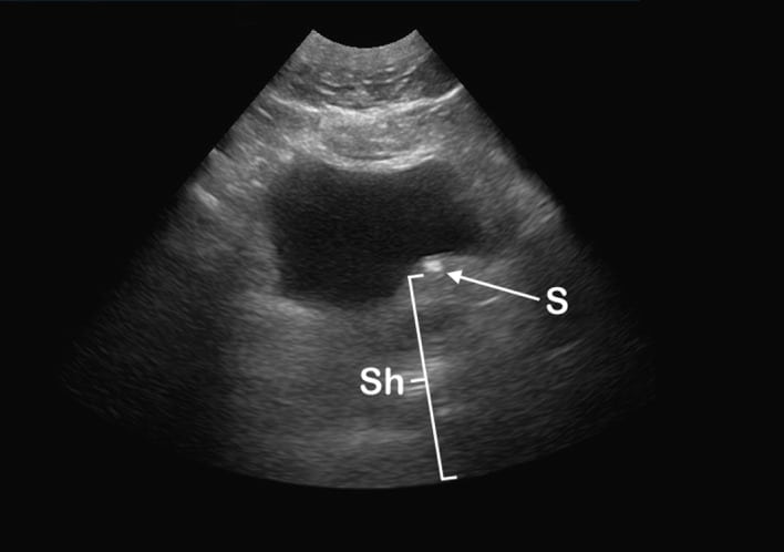
Bladder Bulge Unifying Old And New Sonographic Bladder Wall Abnormalities In Ureterolithiasis The Western Journal Of Emergency Medicine

Ultrasound Tutorial Kidney Bladder Urinary Tract Radiology Nation Youtube
![]()
Efast Ultrasound Exam Made Easy Step By Step Guide Pocus 101

Figure Bladder Dimensions In Sagittal And Statpearls Ncbi Bookshelf
The Radiology Assistant Normal Values In Ultrasound
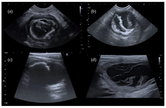
Veterinary Sciences Free Full Text Pseudomembranous Cystitis An Uncommon Ultrasound Appearance Of Cystitis In Cats And Dogs Html
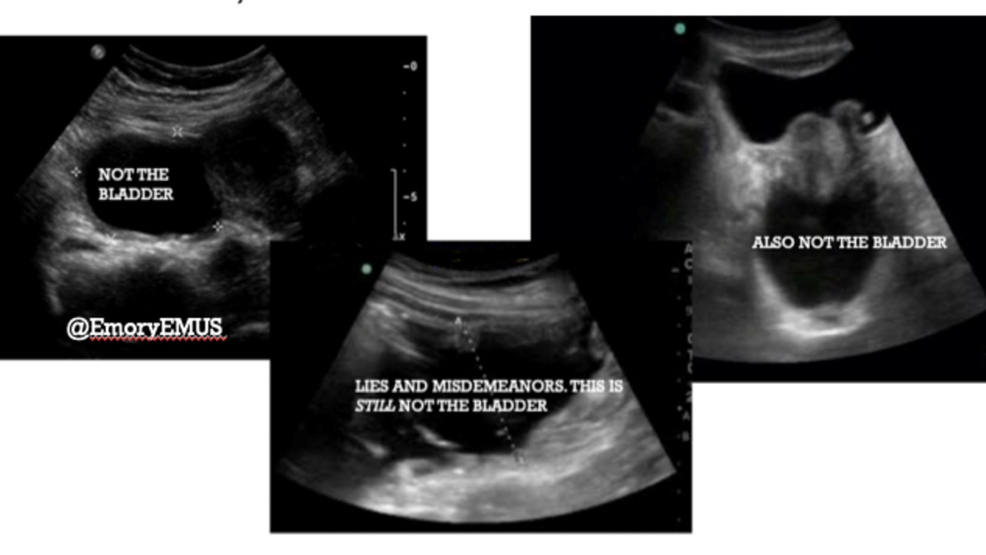
Not So Fast Part 3 Bladder And Pelvis Emory School Of Medicine

Small Animal Abdominal Ultrasonography The Urinary Tract Urinary Bladder Urethra Today S Veterinary Practice

The Ultrasound Bladder Prep Ultrasound Unwrapped

The Radiology Assistant Normal Values In Ultrasound
![]()
Efast Ultrasound Exam Made Easy Step By Step Guide Pocus 101
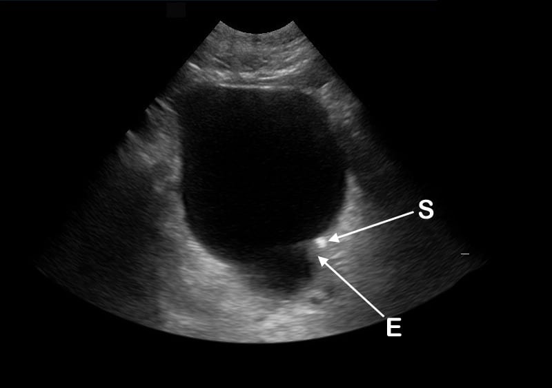
Correlating The Sonographic Finding Of Echogenic Debris In The Bladder Lumen With Urinalysis Cheng 2016 Journal Of Ultrasound In Medicine Wiley Online Library

Top Panel Ultrasound Scan Of Urinary Bladder Of Patient 3 Was Download Scientific Diagram
Bladder Bulge Unifying Old And New Sonographic Bladder Wall Abnormalities In Ureterolithiasis The Western Journal Of Emergency Medicine
Efast Ultrasound Exam Made Easy Step By Step Guide Pocus 101

Small Animal Abdominal Ultrasonography The Urinary Tract Urinary Bladder Urethra Today S Veterinary Practice

2d Gray Scale Ultrasound Image The Maternal Bladder Is Full The Download Scientific Diagram
Post a Comment for "How To Read Bladder Ultrasound"