How To Scan Baby Hips Ultrasound
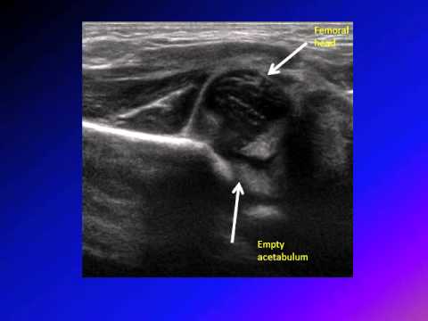
Clinical examination while useful has been shown to be insufficient as the sole screening method in infants. The focus is set at the acetabular edge.
Ultrasonography
The purpose of this study is to report US results and follow-up of.

How to scan baby hips ultrasound. Scanning at greater than 3 months reduces visibility of anatomy. Lower limb of the ilium at the depth of acetabular fossa the midpoint of acetabular roof and the labrum. The coronal view can be obtained with the hip in either the physiologic neutral position 15-20 flexion or the flexed position.
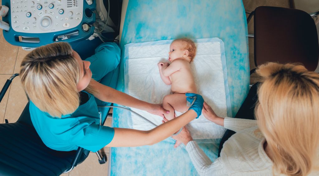
Ultrasound for neonatal clinic. Neonatal Hip UltrasoundDevelopmental Dysplasia of the Hip is a congenital disorder in which the acetabulum is underdeveloped or there is dislocation of the h. To help reassure parents involve them in positioning and comforting the Baby during the scan.
Your child will be placed on their side or back on an ultrasound bed and their knees will usually be bent during the scan. To show the efficiency of hip sonography in detection of developmental dysplasia of the hips in those without clinically. Some babies become restless during the procedure but you will be able to stay with your baby throughout.
Ultrasound examination at 8 weeks in high risk infants is an integral part of the screening process in some units. The Hip Ultrasound Scan. The Barlow manoeuvre test attempts to.
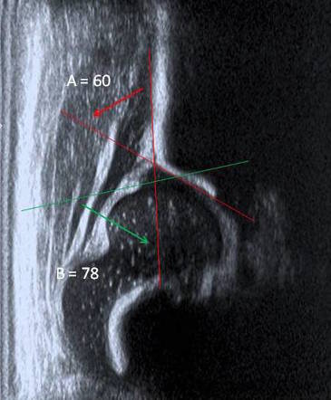
Ultrasonography of the infant hip in the coronal plane has three landmarks. The babys hip must be dynamically scanned with coronal and transverse evaluation with the hip in. There have been childhood hip problems in your family parents brothers or sisters.
Baby having a hip ultrasound at. 1 a straight iliac line. A baby positioning device to assist in keeping the baby in decubitus position should be used.
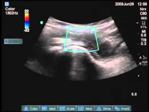
A lineair high frequency probe is used. Important Only an ultrasound examination of the hips enables a detailed diagnosis of a problem in the babys hip joint. Some warm gel will be placed on each hip in turn.
The ultrasound probe is placed on the skin and gently moved around to obtain the best picture of the hip joint. Around 6 months of age enough bone is present in an infant hip to make an X-ray more accurate than ultrasound. You will be able to stay with your baby throughout the scan.
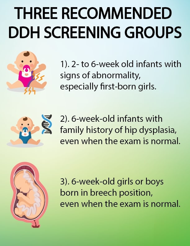
The examiner has to check the classical shape of chondro-osseous junction to. Examination of the Baby. Because of the risk of developmental dysplasia of the hip in infants born breech-despite a normal physical exam-the American Academy of Pediatrics AAP guidelines recommend ultrasound US hip imaging at 6 weeks of age for breech females and optional imaging for breech males.
Tilted probes or obliques scanning may lead to overdiagnoses. For babies who attend for ultrasound scan of the hips after screen positive newborn hip referral standard 4 requires an outcome decision to be made within the target timescale. There is no preparation for this type of ultrasound scan.
You will be asked to partly undress your baby. 2 the tip of the acetabular labrum. An ultrasound scan has been recommended to check your babys hips This leaflet will explain the procedure.
Congenital Hip Dysplasia Dislocation. Perform hip examination should be performed within the first 3-5 days of life prior to hospital discharge. Optimum baby age 6 weeks old.
The scan is a painless procedure. Lie the baby supine Have the babys feet facing you. Hip and groin pain is very common and ultrasound has been proven to be a useful tool in the assessment of the hip tendons ligaments muscles nerves synovial recesses articular cartilage bone surfaces and joint capsule.
Your baby should have an ultrasound scan of their hip between 4 and 6 weeks old if a doctor midwife or nurse thinks their hip feels unstable. Your baby will be placed on hisher side in a special cradle whilst the pictures are being taken. Some gel will be placed on each hip in turn.
Flexion with and without stress. Next the transducer is moved backwards and forwards from the basic position to identify the round structure of the hip joint. It is important to display an image in the coronal plane at the level of the triradiate cartilage which is the synchondrosis between the iliac ischial and pubic bones which form the acetabulum.
The ultrasound transducer is then placed in the anatomic coronal plane Fig. Ultrasound linear transducer is placed parallel to the lateral aspect of the infants hip. Use 5-75 or more MHz linear probes no trapezoid or sector probes.
Perform Barlow and Ortolani manoeuvres on each hip. Any child of any age with an Ortolani or Barlow sign should be referred to a pediatric orthopedist for evaluation. There are numerous ways that you can scan the baby.
If I suspect DDH in a baby when should I refer to a pediatric orthopedist. Three landmarks should be well visualized on a sonogram. Early detection shortens the time of treatment and also means a good chance of avoiding an operation later in life.
Examine the hips by gently abducting and adducting each hip. And 3 the transition from the os ilium to the triradiate cartilage. You will be taken into the ultrasound room and introduced to the Sonographer performing the scan.
The goal of the ultrasound scan is to detect and localize pathological processes to differentiate between intra-articular and extra. Clear gel is put on the area to be imaged and a transducer a small smooth handheld device is placed on the hip. Babies should also have an ultrasound scan of their hip between 4 and 6 weeks old if.
Graf Method For Ultrasound Classification Of Developmental Dysplasia Of The Hip Radiology Reference Article Radiopaedia Org

Ultrasound International Hip Dysplasia Institute

Hip Ultrasound Youtube

Ultrasound Of The Infant Hip With Developmental Dysplasia Ppt Video Online Download

Mackenzie S Story Part 1 The Diagnosis Hipsleepers
![]()
Ultrasound Of The Infant Hip With Developmental Dysplasia Ppt Video Online Download
St How To Hip Ultrasound Exam Youtube
Hip Dysplasia Should My Child Be Screened Uva Radiology
An Index For Diagnosing Infant Hip Dysplasia Using 3 D Ultrasound The Acetabular Contact Angle Semantic Scholar
![]()
Infant Hip Ultrasound Developmental Dysplasia Of The Hip 6 22 17 Ppt Download
Ddh Developmental Dysplasia Of Hip Congenital Hip Dislocation Chd Youtube
Ultrasound Infant Hip For Parents Hackensackumc

Reliability Of 2d And 3d Ultrasound For Infant Hip Dysplasia In The Hands Of Novice Users Semantic Scholar

Standard Coronal Ultrasound Section Through The Acetabulum Of A Normal Download Scientific Diagram

Normal Hip Us Scan Of 6w Post Natal Infant Coronal Right And Axial Download Scientific Diagram
Hip Dysplasia Should My Child Be Screened Uva Radiology

6w Postnatal Scan Axial Right And Coronal Left Us Scans Of A Download Scientific Diagram

Normal Hip Us Scan Of 6w Post Natal Infant Coronal Right And Axial Download Scientific Diagram

Neonatal Hip Ultrasound Ultrasound Diagnostic Medical Sonography Ultrasound Sonography
Post a Comment for "How To Scan Baby Hips Ultrasound"