4d Ultrasound Of Baby With Down Syndrome
This can cause mental impairment and physical abnormalities. The ultrasound examination cannot diagnose a fetus with Down syndrome with certainty.

3d 3rd Trimester Ob Ultrasound Images 3d 4d Ultrasound Performed In The Early Third Trimester Usually Provide Images That Look More Like A Baby And Less Li
The blood test which you may have any time after 10 weeks or on the same day as your scan is.

4d ultrasound of baby with down syndrome. An ultrasound can detect fluid at the back of a fetuss neck which sometimes indicates Down syndrome. Down syndrome is the most commonly occurring chromosomal abnormality during pregnancy. Ultrasonography should not be used by itself to diagnose or exclude Down Syndrome.
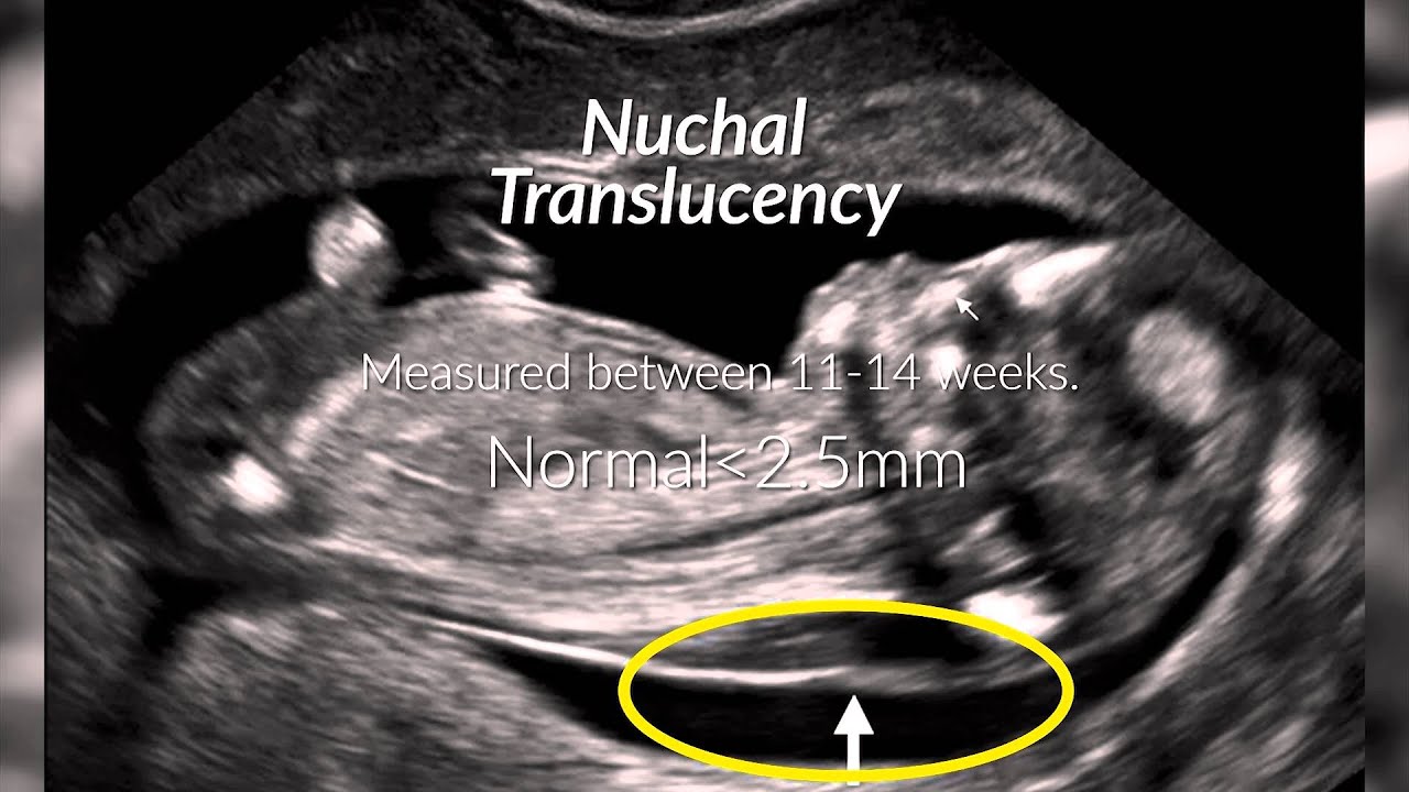
The First Trimester Screen with Nuchal translucency evaluation is a non-invasive evaluation that combines a blood test drawn on the mother at approximately 11 weeks with a very specialized ultrasound of the back of your babys neck between 11-14 weeks gestation to identify your level of risk for having a baby with. Babys with Down Syndrome have an increased amount of fluid. Ultrasound in Obstetrics and Gynecology Meta-Analysis of Second-Trimester Markers for Trisomy 21 January 2013.
Ultrasound in Obstetrics and Gynecology Isolated fetal pyelectasis and the risk of Down syndrome. Cell-free DNA has a detection rate for. A meta-analysis December 2013.
The answer to that question is yes. Pediatrics 31 years experience. What causes Down syndrome in a child.
These printed and digital custom fake ultrasounds look very close to the real deal. He also measured and studied the babys nose. Soft markers are sonographic findings that do not in themselves cause any adverse outcomes.
Ultrasound of baby with down syndrome. Two-dimensional ultrasound images of fetal profile FP line at. It occurs when there is an extra copy of chromosome 21.
Ultrasound waves cause no harm to the mother or fetus. The ultrasound examination cannot diagnose a fetus with down syndrome with certainty. Hole in the heart no bone in the small fingers and the length of the babys middle toe.
Theres nothing like the excitement of seeing your baby for the first time. Baby face 4d ultrasound indianapolis indiana. It affects about 1 in 700 babies.
There is a group on facebook to support moms who are pregnant and have been told they have an EIF. This fetus has Down syndrome with a ventricular and atrial septal defect. I had a 4d scan at around 20 weeks but it was specifically what they call a fetal abnormality scan where the obstetrician looked for physical signs of downs syndrome in the baby eg.
How to tell if a baby is head down. 4D scans are 3D images of your baby while the baby is moving. These scans are used during the course of the pregnancy to monitor fetal growth and development.
This is an effective method in the early detection of health disorders. Im already getting a 4D ultrasound in January so theyre using that method. During the first trimester this combined method results in more effective or comparable detection rates than methods used during the second trimester.
A position zero in a euploid fetus at 24 6 weeks gestation. C position positive in a fetus with Down syndrome at 28 2 weeks. First Trimester Screen with Nuchal Translucency Evaluation.
A pregnancy ultrasound or scan is an imaging test that uses sound waves to see how a baby is developing in the womb. An ultrasound test measures nuchal translucency. My health care center called and let me know that the test for Down Syndrome and Spinabifita Im pretty sure came back triggering for Down Syndrome and that I would need further testing to see if my child does have down syndrome.
The ultrasound test is called measurement of nuchal translucency. Adults with Down syndrome may live about 60 years but this can vary. 3D scans are realistic still pictures of your baby and are useful in helping doctors look for abnormalities.
Sensitivity for detecting Down Syndrome is increased when ultrasound findings are interpreted in combination with serum analyte screening tests such as first and second trimester screening and integrated and sequential screening. Certain findings sometimes called soft markers on ultrasound may make your doctor more suspicious that your baby may have Down syndrome. EIFs were considered a soft marker for down syndrome but now they are showing up more and more now that there are more advanced ultrasound machines.
Ultrasound can detect fluid at the back of a fetus neck which can be an indicator of down syndrome. Sometimes a doctor will order an ultrasound as an additional possible indicator when other factors such as a maternal serum screening indicate an increased possibility of Down. I wasnt overly concerned the ultrasound lady said it was normal and they.
A 46-year-old member asked. An increased desire to and the medical possibility of learning of fetal anomalies during the first trimester has led to great interest in using ultrasound to determine if a fetus has Down syndrome. And d position negative in a trisomy-18 fetus at 23 5.
The egg and sperm cells then divide in half. Will You Be Able to Tell If A Baby Has Down Syndrome in an Ultrasound. B position zero in a fetus with Down syndrome at 21 3 weeks.
Although there is no way of preventing Down syndrome there are signs in pregnancy that can determine if a baby has it. Testing for Down syndrome During the ultrasound scan the sonographer measures the fluid beneath the skin at the back of your babys neck - known as the nuchal translucencyThis measurement is combined with other information to estimate the risk of Down syndrome. Approximately 30 of babies with Down syndrome have detectable abnormalities on the mid-trimester ultrasound 1.
This normal accumulation of fluid is known as the Nuchal translucency NT measurements. When a baby is conceived a normal egg cell and normal sperm cell start with 46 chromosomes. However ultrasound is often used as a screening test for Down syndrome and other chromosome abnormalities.
My Dr told me not to worry about it at all. The NT ultrasound examination involves measuring the amount of fluid on the back of the babys neck. These examples illustrate pathology observed in fetuses with Down syndrome.
Ultrasound cannot diagnose a fetus with Down syndrome trisomy 21. However they are seen more frequently in fetuses with an abnormality. Down syndrome is one of the most common genetic birth defects.
It can pick up soft markers for downs. Three and 4D ultrasound allows the physician to simultaneously examine multiple views of the heart and vessels as well as evaluate the heart in a 3D and 4D model. When can you find out your baby has down syndrome through an ultrasound.

Pin On Baby Development Week By Week

Accuscan Health Imaging Utah 3d 4d Ultrasound

Pin On Down Syndrome

12 Week 4d Scan Circulatory And Digestive Systems Pancreas And Liver Begin Early Functions 13 Weeks Pregnant Baby Breastfeeding 12 Week Ultrasound Pictures

Amniotic Band Syndrome Abs Ultrasound Obstetric Ultrasound Ultrasound Sonography

Before After Photo Gallery 3d And 4d Ultrasound Virginia Baby Ultrasound Pictures Baby Pictures 3d Ultrasound
Pin On Pikatallennukset
Pin On Ob

Pin On Stuff

Pin On Pregnant

Pin En 3d And 4d Ultrasound

3d 4d Ultrasound Baby Scan Window To The Womb Ltd Baby Scan 4d Ultrasound Baby Ultrasound

Pin On Places To Go For Ultrasound Scan

Before After 2008 4d Ultrasound Ultrasound 3d Ultrasound

20 Week Ultrasound Why Is It Important Baby Ultrasound Pictures Ultrasound Pictures 20 Weeks Pregnant Ultrasound

3d 4d Ultrasound Baby Scan Window To The Womb Ltd Baby Ultrasound Girl Ultrasound Pictures 4d Ultrasound

Pin On Ultrasound Notes

Ultrasound Atlas Glowm Medical Ultrasound Obstetric Ultrasound Ultrasound

Pin On Obstetrică Patologica
Post a Comment for "4d Ultrasound Of Baby With Down Syndrome"