When Can You Do An External Ultrasound
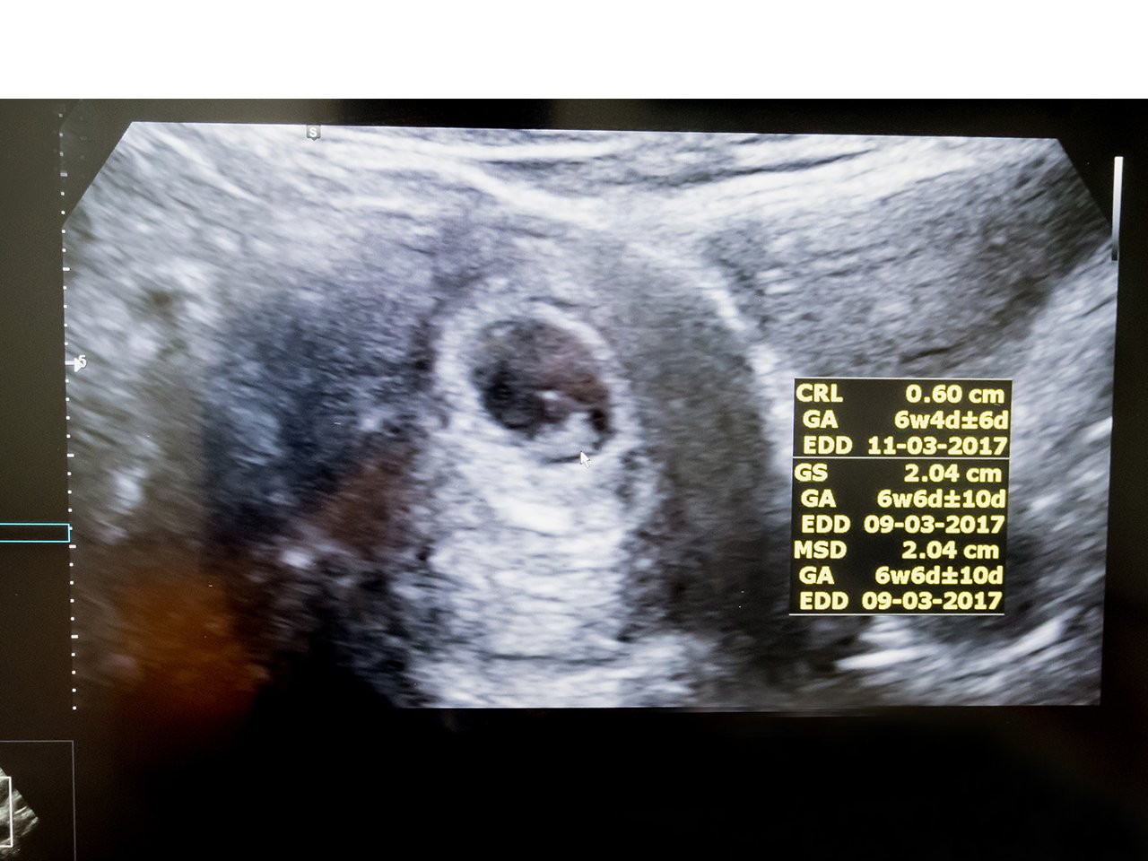
Exactly when you get your first ultrasound exam may vary however if you have certain chronic conditions including asthma or obesity. In viable pregnancies trans-vaginal internal scans should be able to detect a gestation sac from 5 weeks of pregnancy.
Pregnancy Ultrasound When To Get Your First Ultrasound

From this point in a pregnancy internal scans are usually only performed if an external scan cant produce a clear image.

When can you do an external ultrasound. You may also receive an additional ultrasound in the first trimester before your 14th week of pregnancy. You might have an external ultrasound of your lower tummy pelvis or a vaginal ultrasound to help diagnose ovarian cancer. A transvaginal ultrasound also called an endovaginal ultrasound is a type of pelvic ultrasound used by doctors to examine female reproductive organs.
Ultrasound scans use high frequency sound waves to create a picture of a part of the body. Digital and TVU examinations of CL every 2 weeks from 14 to 30 weeks gestation with examiners blinded to the results of the alternate technique 12 both independently predict PTB but TVU has a much stronger association with PTB than manual examination of the cervix. Trans-abdominal external scan may be less accurate at this early 5 week stage.
It can also be used to examine the liver kidneys and other organs in the tummy and pelvis as well as other organs or tissues that can be assessed through the skin such as muscles and joints. It kind of depends on what they can see. External ultrasound scan.
Ultrasound is also used very frequently during a prostate biopsy to guide the physician to biopsy exactly where needed. Fibroid tumors benign growths masses cysts and other types of. Find out why a doctor might order this type.
Therapeutic ultrasound is a treatment modality commonly used in physical therapy. An ultrasound can help with the diagnosis of multiple conditions related to your tissues or organs. In your ninth week of pregnancy your doctor will recommend an ultrasound to know the size of your growing baby.
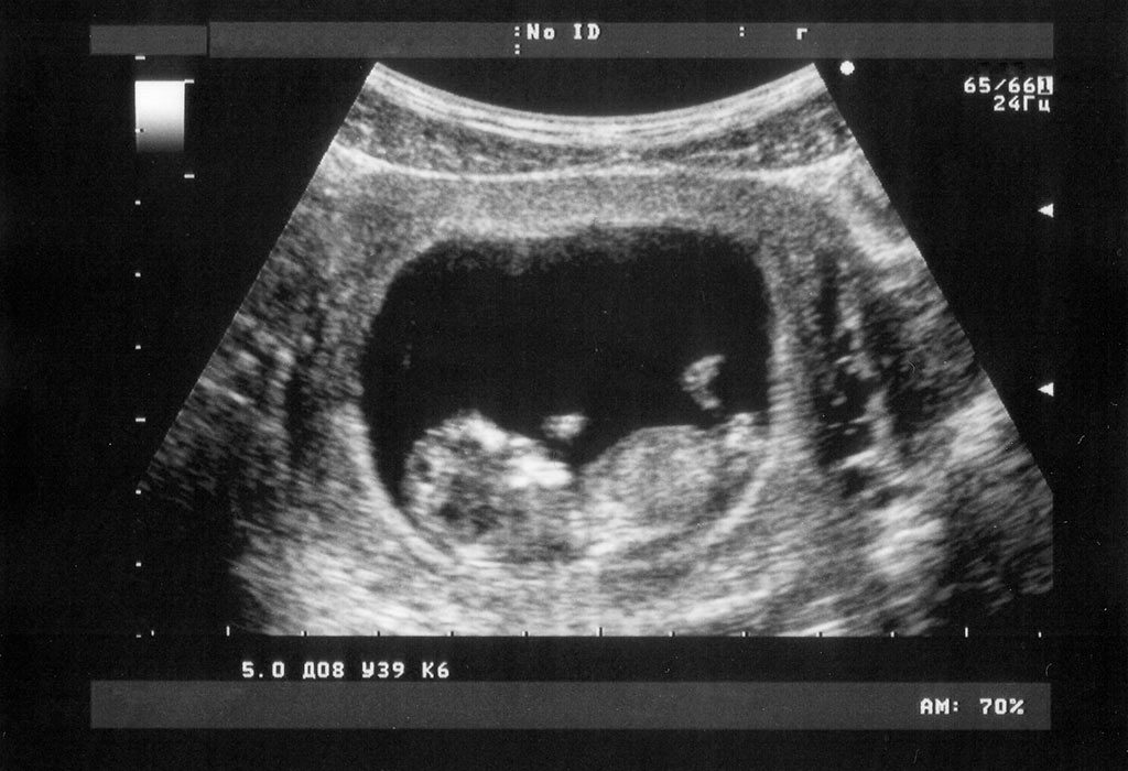
An external ultrasound scan is most often used to examine your heart or an unborn baby in your womb. In the ultrasound scan you will get to see a tiny blob with a heartbeat in your uterus which will eventually grow to become your baby. The type you need depends on the area of your body youre having scanned.
A pelvic ultrasound may be used to diagnose and assist in the treatment of the following conditions. External ultrasound scan - when the doctor or sonographer moves a probe over your skin. Pictures of 8 Week Ultrasound.
You can see little legs and arms the head which is much bigger than the body and the space in which your baby is floating around. Heres what to expect. This new strategy shows promise but also has shown a potential for deleterious side effects.
A yolk sac can be seen at 5 12 weeks gestation. There are different types of ultrasound scans. It sounds less than fun.
It is the first good look you will get of your baby. Instead youll need a transvaginal ultrasound. For instance the Nuchal Translucency Scan performed at 12 to 14 weeks is an external scan.
Internal ultrasound scan - when the doctor or sonographer inserts a probe into your body eg into your vagina or back passage. It is used to provide deep heating to soft tissues in the body. They can also evaluate the status of a patients bones.
These tissues include muscles tendons joints and ligaments. Read on to know more about 9-week ultrasound scan. The ultrasound scan is a staple part of antenatal care across the globe.
An ultrasound can help your doctor determine if your prostate size increase is normal and age-related or a sign of prostate cancer. An external ultrasound scan is most often used to examine the heart or an unborn baby in the womb. Thanks for the responses ladies.
In some cases increased blood flow can indicate an infection. They do an interval until about 12 weeks. Ultrasound evaluation of the gastrointestinal tract should also be the method of choice in patients with severe symptoms when colonoscopy is contraindicated or in cases when a fast examination is needed and access to other.
I had an external one at 4weeks3days to see if i was further along but they couldnt see anything at all so they said i. The quality of ultrasound images produced by both trans-vaginal and trans-abdominal scans will be affected by your BMI and the. In most cases your baby is too small to be seen clearly or at all on an external abdominal ultrasound.
Abnormalities in the anatomic structure of the uterus including endometrial conditions. Certain types of ultrasounds can capture a patients blood flow. It can show the ovaries womb and surrounding structures.
As you can see from this picture the babys body is actually starting to look like a human. Ultrasound in physical therapy is not to be confused with diagnostic ultrasound which is an ultrasound that is used to see the inside of. It can also be used to examine the liver kidneys and other organs in the tummy and pelvis as well as other organs or tissues that can be assessed through the skin such as.
At that point theyll probably try external first and then if they dont get a good view have you pee full bladder for external ultrasounds and do a transvaginal internal one. Direct sonothrombolysis using external typically low frequency ultrasound has been tested for treatment of thrombotic disease such as stroke Siegal and Luo 2008. Ultrasound technology allows for the safe and effective imaging of a developing embryofoetus from the beginning to the end of a pregnancy allowing for your peace of mind and for a close watch to be kept on your health and that of your unborn baby.
Doctors use ultrasounds to diagnose conditions such as.

Pin On First Time Mommy

Ultrasound Sonogram Baby S Feet By Sweetmango On Creativemarket Baby Feet Abstract Artwork Artwork

20 Week Ultrasound Why Is It Important Baby Ultrasound Pictures Ultrasound Pictures 20 Weeks Pregnant Ultrasound

Boy Or Girl Need Ur Help Boy Or Girl Celestial Outdoor
Pin On Baby Bump Breastfeeding Baby Wearing

Pin On Ob Gyn Ultrasound Board
:focal(512x385:513x386)/https://tf-cmsv2-smithsonianmag-media.s3.amazonaws.com/filer_public/90/0b/900b6422-dbec-4369-a3e1-c21ca7bc8d5f/gettyimages-1219040940.jpg)
A Brief History Of The Sonogram Innovation Smithsonian Magazine
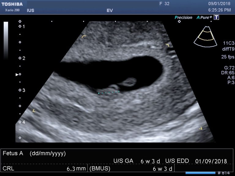
What To Expect At Your Six Week Ultrasound Appointment

Pitfalls In Transvaginal Ultrasound Transvaginal Ultrasound Ultrasound Start Tv

Normal 13 Week Baby Ultrasound Ultrasoundfeminsider Ultrasound Pictures 8 Weeks Ultrasound 8 Week Ultrasound

Nub Theory For Detecting Gender As Early As 10 Weeks Baby Gender Predictor Baby Gender Ultrasound Baby Gender Prediction
6 Week Pregnancy Scan 6 Week Ultrasound 6 Week Scan 6 Week Scan Photo International Ultrasound Services
When You Can See A Baby On The Ultrasound Scan

Pin On Things I Loved As A New Mom
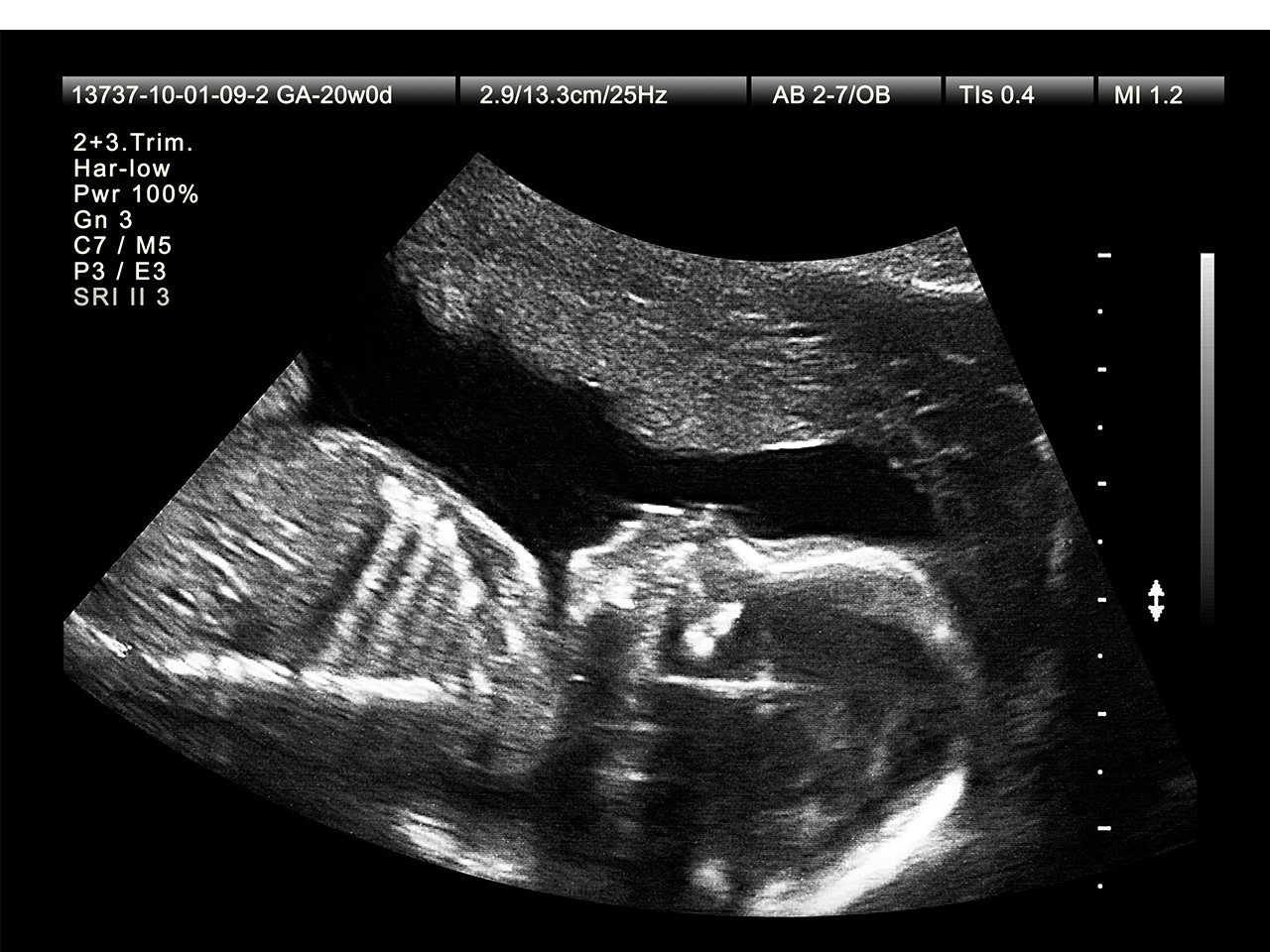
What To Expect At Your 20 Week Ultrasound Appointment

Pin On Baby After Infertility

Ultrasound Imaging Of Hernia Parts 1 2 Of 4 A Youtube Video Tom Wade Md Ultrasound Medical Ultrasound Diagnostic Medical Sonography
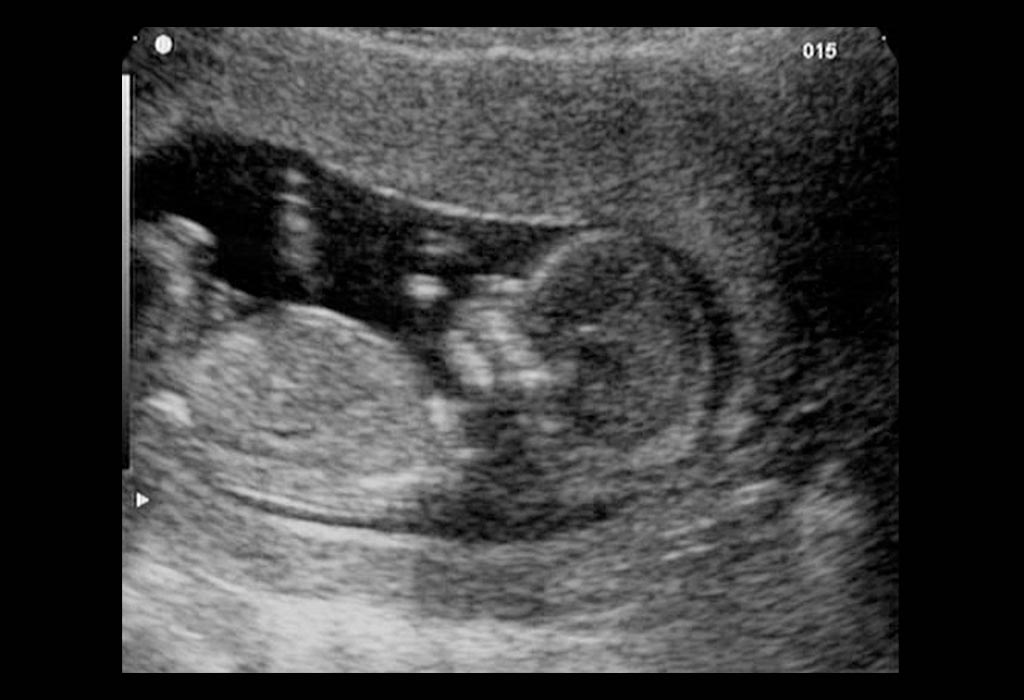
12 Weeks Pregnant Ultrasound Process Abnormalities Accuracy

In Another Case The Transvaginal Ultrasound Shows The Tip Of The Iceberg Sign Acoustic Shadowing From The Dermoid Cyst Ovarian Cyst Transvaginal Ultrasound
Post a Comment for "When Can You Do An External Ultrasound"