When Can You See Baby's Brain On Ultrasound
On our 18 week ultrasound the doctor diagnosed a single cyst on the babys brain choroid plexus cyst. My bloodwork was phenomonally GOOD - 1 in 10000 chance for Trisomy 18 and 1.

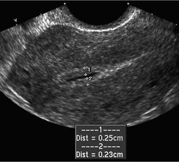
Pin On Precho Babyechografie Antwerpen
Anterior Horns of the Lateral Ventricles.

When can you see baby's brain on ultrasound. I read on another post recently that someone had a similar issue with their 20 weeks scan. The shape of the cerebellum the part of the brain that controls muscle coordination and balance is important in the detection of neural tube defects. I just want my baby to be OK I repeated over and over again on a Thursday morning last AprilThree weeks earlier a sonographer had seen an abnormality in my unborn daughters brain.
Your doctor may recommend more frequent ultrasounds if you have existing health conditions. The neural tube forms in the first few weeks of pregnancy. A transcranial Doppler ultrasound evaluates blood flow in the brains major arteries.
How to see ultrasound of babyWhen you go in for your ultrasound anatomy scan aka the first time you see your baby developed beyond a gestational sac with a heartbeat you probably have images in your head of a perfect button nose pudgy cheeks and a sweet smile just like how she might look. The neonatal head ultrasound is taken by angling transducer back. This is the earliest a heartbeat will be detected.
Ad Discover these surprising facts about ultrasound imaging right now. This image is usually taken during a standard ultrasound. It is most commonly performed on infants whose skulls have not completely formed.
Here is an image taken through the frontal lobes. One umbilical vein that delivers nutrients and oxygen-rich. Click to see full answer Similarly it is asked can you see the brain on an ultrasound.
If your baby was born more than 3 weeks before your due date the doctor will give them a head ultrasound. Cranial UltrasoundHead UltrasoundUltrasound imaging of the head uses sound waves to produce pictures of the brain and cerebrospinal fluid. Ultrasound is safe noninvasive and does not use ionizing radiation.
The umbilical cord typically has three blood vessels. Our specialists are able to confirm this diagnosis with a fetal magnetic resonance imaging MRI exam which provides more detailed images of the brain. Hydrocephalus is typically detected through a prenatal ultrasound between 15 and 35 weeks gestation.
The test checks for brain problems that. An ultrasound of the brain and head is used on infants to evaluate an enlarged head and to look for infection or abnormal growths. The babys heartbeat can be picked up by ultrasound from around 7 weeks so you will actually be able to listen to it during the scan.
Usually the ultrasound images are very clear and you will be able to see most of your babys features by the 27 28th week of your pregnancy. If you have a scan before 7 weeks you wont be able to see much at all. A cranial ultrasound cannot be done until the.
Single umbilical artery This one really freaks people out because the umbilical cord is seen as the babys lifeline but this defect is common and occurs in as many as 1 percent of single-baby pregnancies and almost 5 percent of multiple pregnancies. Cant see baby on ultrasound. Most fetuses 369 65 of 570 who had iuMRI within 2 weeks were less than 24 weeks gestation.
Typically speaking the OBGYN will order one around the middle of the second trimester usually between weeks 16 to 20 to check the babys measurements and screen for any problems. You can see the CSF in the lateral ventricles as a dark image. Fetuses with identified intracranial abnormalities on ultrasound underwent iuMRI mostly within 2 weeks of the ultrasound scan.
Ultrasound imaging of the head uses sound waves to produce pictures of the brain and cerebrospinal fluid. The top of this tube becomes the babys brain and the rest becomes the spinal cord. I cant remember if it was specifically the brain or if it was other parts of the body but the overwhelming consensus was that sometimes that happens.
A brain and head ultrasound is often used to diagnose complications of premature birth including bleeding in the brain. It is possible to see the orbital ridge in the image that forms the inferior boundary. However the small features of the baby can only be seen around 15 weeks.
21 answers last post. Can you see hydrocephalus on an ultrasound. If you have cvs at 10 weeks the results will reveal your babys sex by 12 weeks.
It is most commonly performed on infants whose skulls have not completely formed. When Will You Be Able to Recognise What You re Seeing. The first trimester is months one two and three of your pregnancy.
An ultrasound of the brain and head is used to detect a brain mass in an adult. While most women can expect to see something in a 5-week ultrasound no two pregnancies are the same. Couldnt see brain clearly 20 week scan.
The scans dont come out clear or the baby isnt cooperating. While modern ultrasound technology is relatively reliable a scan that shows the all-clear sign doesnt necessarily mean that everything is okay. An interesting component of the design is that radiologists doing iuMRI were not masked to the ultrasound diagnosis.
14 placenta previa early in the pregnancy during routine ultrasounds many women are told their placentas are low lying.

Placental Abruption Medical Ultrasound Obstetric Ultrasound Ultrasound Sonography
Pin On Ultrasound

Wk 5 Testes Seminoma Diagnostic Medical Sonography Ultrasound Radiology


Pin On Bengal Cat

Gender Baby S Legs Closed Boy Or Girl 20 Week Ultrasound See The Rest Of This Pos Ultrasound Gender Ultrasound Gender Prediction Ultrasound Boy Or Girl

Fnu Offers The 7 Things You Should Know Before Enrolling In A Diagnostic Medical Sonography Student Ultrasound Technician School Diagnostic Medical Sonography

Try And Identify The Structures Labelled A F On This Normal Midline Sagittal Cranial Ultrasound Of A Term Infant Answers At Http Ultrasound Sonography Infant

Baby Yawns In Womb 4d Ultrasound Video Footage Brain Development Ultrasound Technician Premature Baby Development

Ramzi Theory Week By Week Baby Gender Pros Ultrasound Gender Prediction Gender Prediction Ramzi Theory

Pin By Christine Marie On Sonography Diagnostic Medical Sonography Ultrasound Sonography Ultrasound

81 Pics That Ll Freak You Out When You See It Can You Find It Optical Illusions Brain Teasers Horses

Sonogram Hydrocephalus In Ultrasound Ultrasound Ultrasound Sonography Diagnostic Medical Sonography

Pin On Mommy Life Someday

Pin On Twin Pics
Endometrial Measurement In The Presence Of Fluid Medical Ultrasound Ultrasound Sonography Obstetric Ultrasound
Coronal And Sagittal Sonographic Images Demonstrating A Grade 1 Hemorrhage At The Caudothalamic Groove Ultrasound Neonatal Resume

Subcapsular Renal Hematoma Medical Ultrasound Ultrasound Sonography Diagnostic Medical Sonography


Pin On Science

Pda Echo Findings Cardiac Sonography Medical Photos Nursing School Survival
Post a Comment for "When Can You See Baby's Brain On Ultrasound"