How Is A 10 Week Ultrasound Performed
Although it doesnt require any preparation the mother should be sufficiently hydrated and not use creams or oils on the abdomen on the day of the study. Why scan at 7 weeks.

Ramzi Theory Week By Week Baby Gender Pros Ultrasound Gender Prediction Gender Prediction Ramzi Theory
Chorion Villus Sampling CVS performed at 11 weeks Miscarriage rate 1.

How is a 10 week ultrasound performed. Your 10 weeks ultrasound At 10 weeks you are likely to have an abdominal test to track the development of the foetus. How it is performed. This gives parents the same type of.
An abdominal ultrasound can outline and identify fetal and maternal structures. By conducting a scan she will be able to identify whether or not your baby is developing normally. How Is A 5-Week Ultrasound Performed.
Dating Ultrasound 10-13 Weeks Those who forgo the six to eight week ultrasound might have a dating ultrasound around weeks 10 to 13 of pregnancy. This test is performed by a radiologist or gynecologist in the office. At 10 weeks pregnant you will be advised to go in for an ultrasound scan that can determine your babys progress and your own health.
An ultrasound is performed at this stage of pregnancy to confirm your due date confirm if you are having singles twins or more and to visualise your babys heart beating. For High Risk Patients we would offer patients the option of Diagnostic testing Invasive Tests. The ultrasound this week will provide a clearer picture of your baby on the 3D 4D screens.
The first-trimester screening test is a test in pregnancy and consists of both a blood test and an ultrasound sonogram test usually done together between 10 weeks and 13 weeks of pregnancy. But there are a few more details that at 10-weeks an ultrasound can reveal. Most pregnancies arent visible with an abdominal ultrasound until about 8 to 10 weeks.
In the 5 th week of gestation ultrasound is carried out in the transvaginal route instead of the abdominal. Optimal conditions include a blood test done between 8 to 10 weeks and an ultrasound performed by someone with the specific training and whose ultrasound examinations are subject to independent quality control. At 10 weeks of pregnancy the foetus weighs about 4gm and measures around 31cm from crown to rump.
Then using ultrasound as a guide the doctor inserts a needle through your belly or through the vagina while doing a. Just as with the other method the ultrasound is not dangerous. Pregnancies without confirmation or revision of gestational age by ultrasound before 22 07 weeks are considered sub-optimally dated.
I meet with the surgeons in a week to schedule surgery. Ultrasound performed by a medically trained sonographer. The sonographer will put gel on your abdomen and move a hand-held device called a transducer back and forward over your skin.
However a transvaginal ultrasound is performed using a probe that is inserted into the vaginal canal. The youngest female fetus whose gender was correctly identified was 11-week-old and the youngest male fetus whose gender was correctly identified was 11 weeks and 1 day old. It is performed with a small wand that is placed in the vagina and pressed against the cervix in order to get a picture from that direction.
Typically you can expect a quick 15-min check for heartbeat abnormalities and organ functioning. Before the abdominal ultrasound is performed the patient should empty bladder. This is called a transvaginal ultrasound.
A transvaginal ultrasound is usually performed by a sonography technician. If ultrasound is performed between 16 07 and 21 67 weeks and the estimated date of delivery by last menstrual period differs by more than ten days the ultrasound-estimated date of delivery should be used. 15 minute ultrasound performed in 2D 3D 4D as early as 13 weeks no later than 16 weeks 100 accuracy.
Kateryna KukotaGetty Images. Most prenatal ultrasound procedures are performed on the surface of the skin using a gel as a conductive medium to aid the quality of the image. Fetal morphology ultrasound scans.
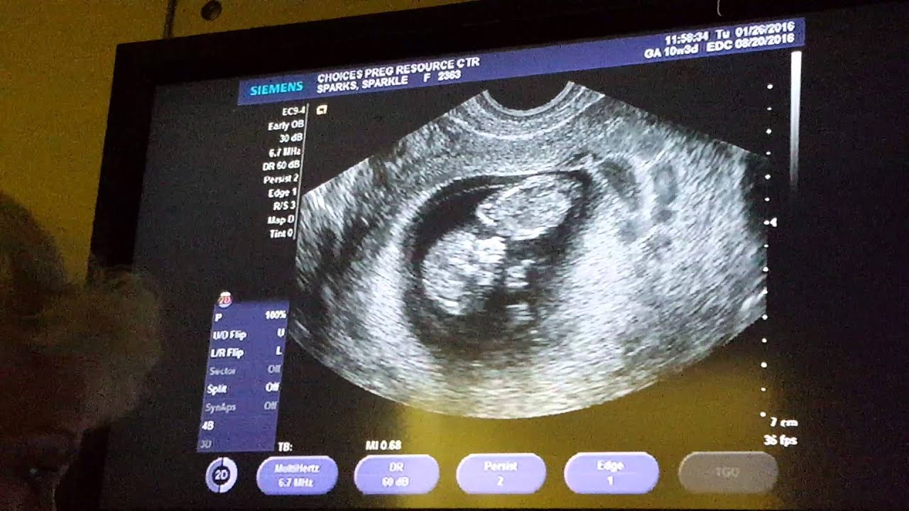
It involves a blood test alone performed at 10-11 weeks The Harmony or Percept Test. Ultrasound image of a fetus at 10 weeks the round area surrounding the baby is called the Amnion this structure is more visible at this time placenta is still developing yolk sac is visible at this time as well your fetus is starting to look more like a baby arms and legs are clearly seen and movements are more noticeable too. Ultrasound imaging was performed in 150 pregnant women 51 34 of whom were in their 11 th week of pregnancy and 99 66 in their 12 th week.
The common first step will be a dating scan between 7-10 weeks as we like to check your baby before 10 weeks. The ultrasound scan gives an accurate estimation of the size of the embryo and the delivery date. Although the blood tests are done previously their results are only taken into account once the ultrasound results are available.
Generally at 20 weeks an abdominal ultrasound can be performed to assess fetal gender. The CVS chorionic villus sampling performed between weeks 10 and 13 uses an ultrasound to determine the placentas location. The World Health Organization WHO mandates at least one ultrasound scan throughout pregnancy.
If the scan is after 10 weeks ultrasound recordings are usually made through your abdomen. Still images and video clips included and saved to a complimentary state-of-the-art USB drive. Listen to your babys heartbeat.
An abnormal first-trimester screening test means there is an increased risk that the fetus has Down syndrome trisomy 21 or another type of aneuploidy. Your doctor may suggest an ultrasound scan at 10 weeks of your pregnancy to check the progress or growth of your baby. Up to 20 cash back Again - a 10 week ultrasound is VERY accurate - you do NOT use the dating scan from 20 weeks IF YOU have a 10 week scan - the EARLIER the ultrasound is done the MORE accurate the scan - the earliest a scan can be done with accuracy is about 6 12 to 7 weeks which is as accurate as 3 to 4 days.
I had an ultrasound a week ago followed by a biopsy four days ago. Amniocentesis performed at 16 weeks Miscarriage rate 05. To do it the pregnant woman lies on a stretcher and uncovers her belly.
My doctor wanted to make sure she performed the ultrasound and biopsy to ensure no cancer cells because if I had cancer and they removed it they may never have known if it had spread.

The Dating Ultrasound At 9 Weeks To Determine The Due Date Of The Baby Ultrasound 9 Week Ultrasound Dating

Pin On Twins

Pin On Baby

Pin On Baby Bumpin Pregnancy

Pin On Baby Development Week By Week

Confirmed Ultrasound Scans A Collection Of Boy And Girl Scans The Gender Experts Ultrasound Gender Ultrasound Gender Prediction Ramzi Theory

Pin On Pregnancy Hacks

The 12 Week Scan When You See Your Real Actual Child For The First Time Is The Most Awe Inspiring Thing You Ll Witness Girl Ultrasound Pictures Ultrasound Pictures 12 Week Scan

Miscellaneous Items And Updates 10 Weeks Pregnant 10 Weeks Pregnant Ultrasound 10 Week Fetus

Nub Theory For Detecting Gender As Early As 10 Weeks Baby Gender Predictor Baby Gender Ultrasound Baby Gender Prediction

9 Weeks Ultrasound 4 Ultrasound 9 Week Ultrasound 9 Weeks Pregnant Ultrasound

14 Weeks Pregnant 2d Ultrasound Active Baby 14 Weeks Pregnant 14 Weeks Pregnant Ultrasound 15 Weeks Pregnant
Pin By Brianna Holzem On Ultrahang Felvetelek Ikrek Twins Ultrasound Baby Ultrasound Pictures Ultrasound

Ultrasounds First Trimester Week 10 10 Weeks 5 Days Amazingpregnancy Pictures Com Trimester By Weeks First Trimester Trimester
Pin On Videos

Pin On Baby By Weeks

3d Image Of A Baby At 10 Weeks Gestation Baby At 10 Weeks Future Baby Portrait Tattoo

10 Week Ultrasound All You Need To Know As A New Mom To Be 10 Week Ultrasound Ultrasound Gender Prediction 10 Weeks Pregnant

Pin On Ultrasound Photos Of Twins Multiples
Post a Comment for "How Is A 10 Week Ultrasound Performed"