Baby Ultrasound Dilated Kidney
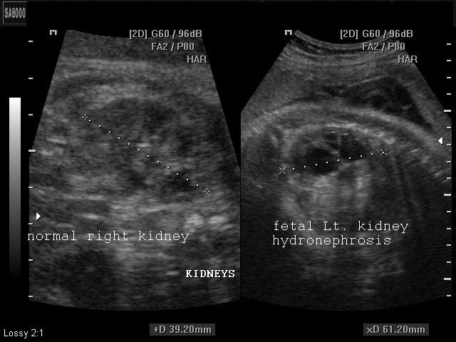
That literally translates to water on the kidneys but it refers to a dilation of the kidney. Because kidney dilation seen at 20 weeks often completely resolves later in pregnancy your doctor or midwife will send you for a follow-up ultrasound at 32-36 weeks to reassess the kidneys bladder and.

Renal Pelvis Dilatation Sciencedirect
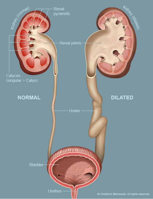
Urine moves out of the kidney and into the bladder through a narrow tube called the ureter.

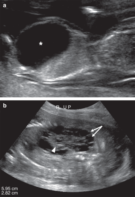
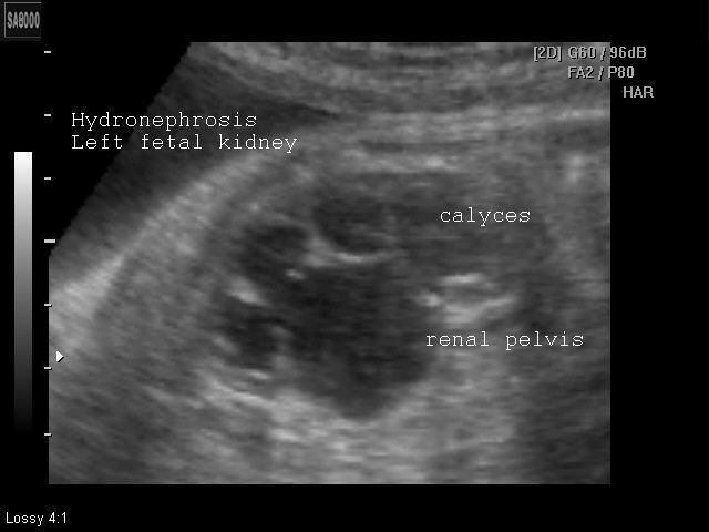
Baby ultrasound dilated kidney. The renal pelvis is where urine collects inside the kidney. Hydronephrosis in newborns is enlargement or dilation of the renal pelvisthe basin in the central part of the kidney where urine collects. Hydronephrosis does not generally cause any long-term problems if its.
I went in for my 20 week utrasound and everything checked out fine but the doctor did say that the baby had some kidney pelvis dilation pyelectasis. Dilation of the renal pelvis can be normal but dilation of the calyces is typically abnormal. A babys kidneys are routinely checked during a second trimester prenatal ultrasound.
When the letter from the consultant pediatrician arrived it said that both kidneys were dilated and were not sure if this was a typo in the. MsLuvN_Motherhood 2 kids. In most cases mild dilation swelling of the kidney is seen on the ultrasound routinely done at the 20 th week of pregnancy.
Close observation with periodic ultrasound to make sure the dilation in the kidney goes away. Unlike others who responded there was no fluid seen on the 20 week ultrasound but enough to be concerning at the 32 week which caused us to go back for another at 36 weeks where it has increased in size. Youngstown Ohio 13 posts.
This occurs in 1 per 100 pregnancies. Ad Check over our 18 symptoms of kidney disease. Causes of hydronephrosis In most cases there are no other abnormalities.
Use of a low dose of antibiotic once a day to prevent infection. So I had my 20wkUltrasound today and my High Risk docJust about scared the hell out of me and my boyfriend by saying everything was normal EXCEPT my son has a enlarged Kidney which is a marker for DS Down SyndromeI have to. Unborn Babies With Dilated Kidneys.
I have read that this is more common in boys because there uterers develop slower than girls sometimes. 1 A prenatal ultrasound ultrasound during pregnancy may reveal a fetus with dilated kidneys. Your Child Has Hydronephrosis.
2 An ultrasound done as a routine evaluation for another medical problem such as a urinary tract infection or incontinence may also reveal hydronephrosis. When dilation of the renal collecting is found on an ultrasound a grading system is used to rate the severity of the hydronephrosis. It is the most common kidney problem found in babies.
Im worried that I have either done something wrong or it could be genetic as my side of. My baby has a dilated left kidney and ureter. Yes dialated kidneys are very common and most go back to normal or work perfectly fine after the baby is born but it is a good idea to keep an eye on it just in case.
The condition affects one in 500 live-born babies and is the most common abnormality detected on prenatal ultrasound scans accounting for up to two-thirds of all prenatally detected defects. It can affect people of any age and is sometimes spotted in unborn babies during routine pregnancy ultrasound scans. Different types of problems can be found including absence of one or both kidneys abnormal position of a kidney hydronephrosis swelling of a kidney fluid-filled cysts and tumors.
The sonographer looks at the size and shape of the kidneys and measures the amount of urine. This is actually quite common occurring in as many as 5 of all pregnancies. Hydronephrosis is a condition where one or both kidneys become stretched and swollen as the result of a build-up of urine inside them.
Baby diagnosed with fetal pyelectasis kidney dilation at 20 week ultrasound. They like the number to be less than 4mm and it was around 41 in both kidneys. Some studies raised concerns about a small risk for Down syndrome with this ultrasound finding.
May 7 2019 at 1155 AM. 20 week Ultrasound reveals Enlarged Kidney-. This is known as antenatal hydronephrosis.
Less often a dilated renal pelvis is an early sign of a more serious problem with the bladder kidney or ureterDown syndrome. So I went for my 18-20wk ultrasound and my unborn son had a slightly dilated left fetal kidney. In some cases hydronephrosis is mild and goes away on its own without treatment.
This condition is often diagnosed before birth during a prenatal ultrasound. When urine is made from the kidneys it first enters a calyx which then empties into the renal pelvis. Has anyone had any experience with this I am 22 weeks pregnant with a baby boy and had a level II ultrasound done 2 weeks ago which showed the babys kidneys slightly dilated they want me to follow up on 6705.
Every child is different and so each treatment plan is tailored to your childs specific needs. With many pregnant women routinely getting ultrasounds their fetuses are more frequently being diagnosed with a condition known as hydronephrosis. Urinary tract dilation is diagnosed by ultrasound.
The renal pelvis is considered large dilated. Hydronephrosis can occur in one or both kidneys. Just so is hydronephrosis a sign of Down syndrome.
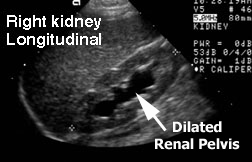
In recent years better ultrasound machines have allowed your doctor to see your babys kidneys more clearly during pregnancy. This baby also has a dilated right kidney and will have an ultraosound soon after birth. 18 Warning Signs Symptoms of Kidney Infection You Should Not Ignore.

Kidneys And Urinary Tract Diagnosis Of Congenital Abnormalities The 18 23 Weeks Scan
My Baby Has Swollen Kidneys What Is Prenatal Hydronephrosis
Echogenic Fetal Kidneys Differential Diagnosis And Postnatal Outcome Iame

Fetal Hydronephrosis Radiology Reference Article Radiopaedia Org
A Gallery Of High Resolution Ultrasound Color Doppler 3d Images Fetal Urogenital
Congenital Urinary Tract Obstruction Defining Markers Of Developmental Kidney Injury Pediatric Research

Kidneys And Urinary Tract Diagnosis Of Congenital Abnormalities The 18 23 Weeks Scan
Kidney Ultrasound Hydronephrosis

Hydronephrosis Pavilion For Women

Ultrasound Appearance Of Utd A2 3 A And B Fetal Kidneys At 20 Weeks Download Scientific Diagram

Fetal Ultrasound At 16 Weeks Of Gestation A Fetal Kidneys With Download Scientific Diagram

Renal Pelvis Dilatation Sciencedirect

Kidneys And Urinary Tract Diagnosis Of Congenital Abnormalities The 18 23 Weeks Scan

Echogenic Fetal Kidneys Differential Diagnosis And Postnatal Outcome Iame
A Gallery Of High Resolution Ultrasound Color Doppler 3d Images Fetal Urogenital

Ultrasound Appearance Of Utd A1 A And B Fetal Kidneys At 19 Weeks Download Scientific Diagram

A Pediatrician S Dilemma Understanding Diagnosing And Treating Antenatal Hydronephrosis

Dilated Renal Pelvis Hkog Info
Urinary Tract Dilatation Midwest Fetal Care Center
Post a Comment for "Baby Ultrasound Dilated Kidney"