Reasons For Fetal Kidney Dilation
Hydronephrosis or urinary tract dilation refers to dilation of the pelvis and calyces of the kidney where urine is collected. Usually the swelling goes away on its own either during the pregnancy or after the baby is born.

My Baby Has Swollen Kidneys What Is Prenatal Hydronephrosis
The likelihood of an underlying abnormality increases with the degree of dilatation.

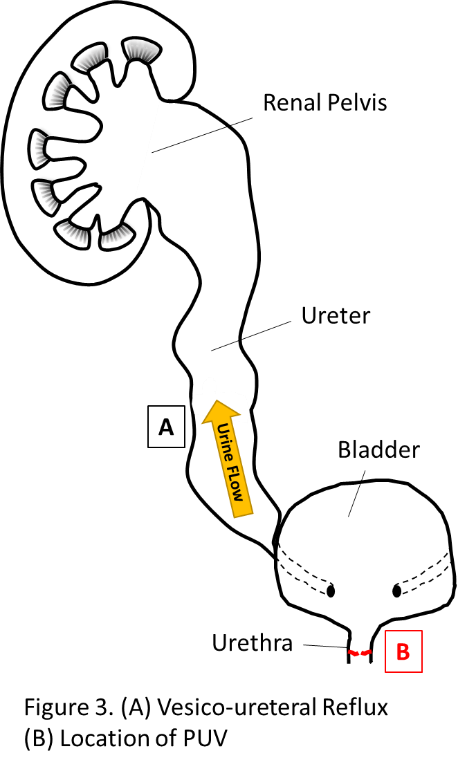
Reasons for fetal kidney dilation. Fifty percent of these babies have a condition called hydronephrosis. The most common underlying abnormalities are ureteropelvic junction obstruction and vesicoureteral reflux. In other cases hydronephrosis is caused by a blockage in the urinary tract or reflux of urine from the bladder to the kidney.
The swelling is often caused by a blockage or narrowing of the urinary tract which stops or slows the urine from leaving the babys body. This is called transient hydronephrosis. Hydronephrosis also occurs when there is a back flow of urine from the ureters into the kidney.
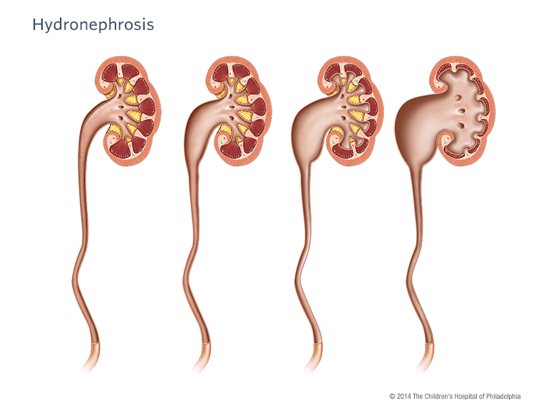
In some fetuses and newborns with hydronephrosis health care professionals cant find a cause and the hydronephrosis goes away on its own. Pictured left to right. Both of these conditions are treatable.
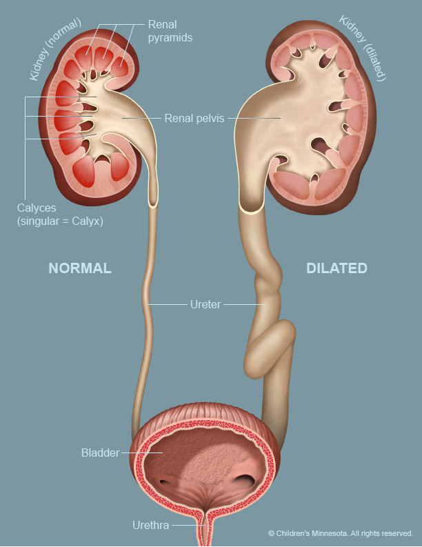
What causes a dilated renal pelvis. The most common reason for fetal kidney dilation is an obstruction of some type. Right kidney appears normal.
Severe RPD 15 mm is frequently associated with urinary tract pathology. Normal appearance of the renal pelvis followed by increasing severity of hydronephrosis. For the majority with mild 5 to 9 mm to moderate 10 to 15 mm RPD however there is uncertainty about the risk of abnormalities and how much postnatal investigation is required.
Here are 16 of the most common symptoms. The Society for Fetal Urology hydronephrosis grading system. Renal pelvis dilatation RPD occurs in 1 of fetuses.
Ad Kidney disease is very serious. Hydronephrosis which may affect how the kidneys continue to develop and function properly during the rest of the pregnancy and after birth. Hydronephrosis occurs when the pelvis becomes enlarged because urine is collecting in the area of the kidneys.
Both ureters are not dilated- left pelviaactatsis featuresdo i need to worry. However sometimes a dilated renal pelvis is due to a block obstruction in the ureter or urine moving back into the kidney reflux. Excess urine collects in the renal pelvis when one or both ureters are blocked restricting the amount of urine that drains out of the kidney.
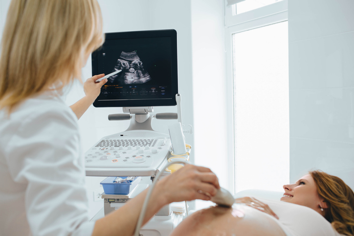
My wife is in 36 th week and she went for scan and it shows fetal left kidney shows isolated renal pelvis dilation measures 8mm. There is wide range in the size of the renal pelvis. With many pregnant women routinely getting ultrasounds their fetuses are more frequently being diagnosed with a condition known as hydronephrosis.
Urinary tract dilation occurs when part of the unborn babys urinary tract swells dilates abnormally with excessive amounts of urine. Fetal renal pelvis dilatation is identified in 1 to 5 of pregnancies and is often a transient normal variant. Often children who have hydronephrosis are born with this condition.
Depending on the extent of the dilation in the area of the kidney where urine is collected is enlarged or dilate. No dilatation calyceal walls are apposed to each other. The grading system of the Society of Fetal Urology SFU classifies hydronephrosis in 4 degrees grade 0-4 and takes into account the degree of pelvic dilatation the number of calyces seen and the presence or severity of renal parenchymal thinning or atrophy Fernbach et al 1993.
Prenatal hydronephrosis which may also be called antenatal. Often children who have hydronephrosis have it from the time of birth. For many babies the large size is just part of the normal range.
Children with reflux are at higher risk for urinary tract infection and may be placed on preventive prophylactic antibiotics at birth. That literally translates to water on the kidneys but it refers to a dilation of the kidney. The amount of amniotic fluidliquor water surrounding the baby in the womb can also be affected if.
Doctors can diagnose hydronephrosis when the enlargement exceeds. This is actually quite common occurring in as many as 5 of all pregnancies.
Prenatal Hydronephrosis Ucsf Department Of Urology

Renal And Kidney Disease Fetal Conditions We Treat Fetal Care Maternal Fetal Care High Risk Obstetrics Ur Medicine Obstetrics Gynecology University Of Rochester Medical Center

Anomalies Of The Tcf2 Gene Are The Main Cause Of Fetal Bilateral Hyperechogenic Kidneys American Society Of Nephrology

Hydronephrosis Pavilion For Women

Ultrasound Appearance Of Utd A2 3 A And B Fetal Kidneys At 20 Weeks Download Scientific Diagram
Hydronephrosis Urinary Tract Dilation Children S Hospital Of Philadelphia
Urinary Tract Dilatation Midwest Fetal Care Center

Ultrasound Appearance Of Utd A1 A And B Fetal Kidneys At 19 Weeks Download Scientific Diagram

Fetal Hydronephrosis Radiology Reference Article Radiopaedia Org

Outcome Of Fetal Renal Pelvic Dilatation Diagnosed During The Third Trimester Wollenberg 2005 Ultrasound In Obstetrics Amp Gynecology Wiley Online Library

My Baby Has Swollen Kidneys What Is Prenatal Hydronephrosis
My Baby Has Swollen Kidneys What Is Prenatal Hydronephrosis

Renal Pelvis Dilatation Sciencedirect

Fetal Hydronephrosis Lurie Children S
Prenatal Hydronephrosis Ucsf Department Of Urology


A Pediatrician S Dilemma Understanding Diagnosing And Treating Antenatal Hydronephrosis

Kidneys And Urinary Tract Diagnosis Of Congenital Abnormalities The 18 23 Weeks Scan
My Baby Has Swollen Kidneys What Is Prenatal Hydronephrosis

Multidisciplinary Consensus On The Classification Of Prenatal And Postnatal Urinary Tract Dilation Utd Classification System Journal Of Pediatric Urology
Post a Comment for "Reasons For Fetal Kidney Dilation"