How To Find Bpd On Ultrasound
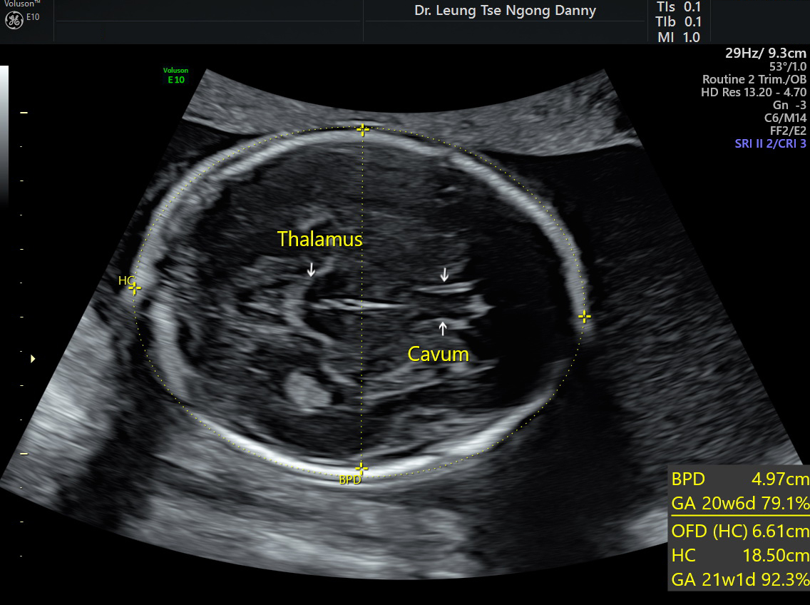
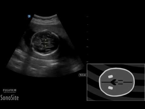
It is a measurement of the diameter of a developing babys skull from one parietal bone to the other. The transducer must be perpendicular to the central axis of the head and thus the hemispheres and calvaria should appear symmetric.

Assessment Of Fetal Gestational Age By Ultrasonic Measurement Of Bi Parietal Diameter In The Southern Part Of Rajasthan
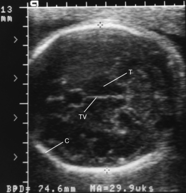
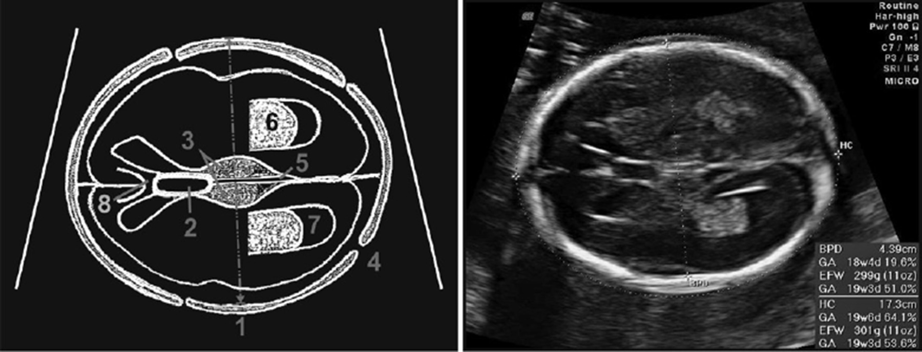
The BPD should be measured on an axial plane that traverses the thalami and cavum septum pellucidum.
How to find bpd on ultrasound. Am J Roentgenol 13783 1981 Sonar -- the Story of an Experiment by Professor Ian Donald which appeared in Ultrasound in Medicine and Biology vol 1 pp109-117 1974. The measurements are prone to both intra- and inter-observer variability. Both of these measurements can be used to confirm gestational age.
Ultrasound technology has demonstrated that biparietal diameter is useful and accurate in determining gestational age of the foetus 5 8. The Hadlock-formula is being widely used for the estimation of fetal weight. Biparietal diameter is used to estimate fetal weight and gestational age.
A linear relationship between growth of fetal femur length FL and biparietal diameter BPD after 22 weeks gestation is described. The first bpd test came back positive on Jan. Gestation by HC is calculated.
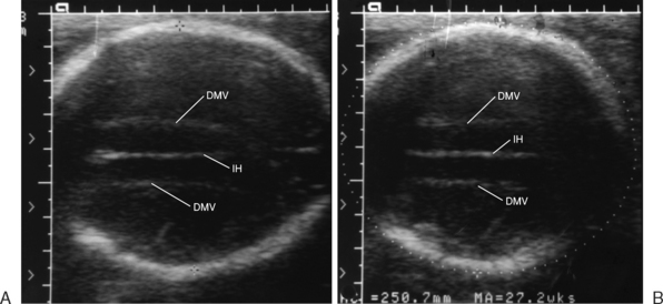
Gestation by BPD is calculated using the formula. The BPD and OFD are measured on a transverse axial section of the fetal head which includes the falx cerebri anteriorly and posteriorly the cavum septum pellucidum anteriorly in the midline and the thalami. Outer edge of the near calvarial wall.
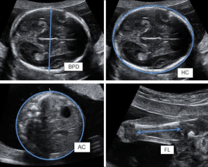
Head Circumference HC measures around the head. The weight of the fetus at any gestation can also be estimated with great accuracy using polynomial equations containing the BPD FL and AC. Days 2 BPD 442.
This will be gestation at time of ultrasound. Biparietal diameter BPD in foetus has been reported to correlate well with gestational age. Gestational age is estimated in weeks from the first day of the LMP.
Youtubersakhi subsribemy other vdoshow to read baby ultrasound anomaly2 d levelhttpsyoutubewcMOu_1OsX4बन अलटरसउड. During an ultrasound your doctor measures the babys head body and thigh bone. Fishel an infectious disease physician at the Cleveland Clinic a bpd blood test is.
Due Date Date of first day of last menstrual period LMP 9 Calendar months 7 days. Abdominal Circumference AC measures around the abdomen. In order to calculate fetal weight you will need the AC and BPD measurement the abdominal circumference and diameter of your babys head.
BPD outer to outer OFD outer to outer HC BPD OFD x 162 HC 1580mm 190 wks. The normal ratio of femur length to BPD FLBPD ratio was found to be 79 - 8. There is an important difference in generating an ultrasound EDD using this methodology from the traditional ultrasound charts.
Enter the measurements in the highlighted box above and then get the weight of your baby. Taking a BPD of say 40 mm the data shows us that on average the woman will deliver a healthy normal baby after 150 days. Fetal biometry measures your babys size.
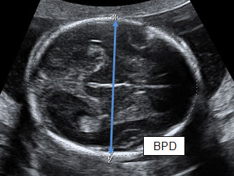
Comparison of ultrasound femur length and biparietal diameter in late pregnancy. Biparietal diameter BPD measures across the head. The BPD measurement is obtained from outer skull bone to inner skull bone leading edge to leading edge perpendicular to the falx at the maximum diameter.
The fetal dating process re-quires the localization of an ultrasound scan plane containing given anatomic landmarks and placement of measurement markers. For example a BPD of 90 cm and an AC of 300 cm will give a weight estimate of 285 kg. The fetal head should occupy at least 30 of the image and should appear as.
HC 162 BPD OFD 3. It helps show your babys development. It is calculated by measuring the maximum width BPD of the cranium divided by its maximum length occipital frontal diameter OFD.
Femur Length FL measures the length of the thigh bone. An estimate of fetal weight EFW can be calculated by combining the above measurements. This estimated fetal weight calculator will calculate percentiles as well as the estimated fetal weights based ultrasound data and on many published formulas.
Every human has two parietal bonesone on the. Inner edge of the far calvarial wall. Effective uses of the FLBPD ratio include its use as a quality control check on.
This will be gestation at time of ultrasound. To calculate the estimated fetal weight four measurements AC BPD HC FL are entered into a mathematic formula. If the available ultrasound equipment allows for measurement of an ellipse this may be drawn around the outside of the calvarium.
Based on a very large population which is quite feasible the confidence interval for this. Weeks eTo1854 0010451 Head_Circumference - 0000029919 Head_Circumference 2 0000000043156 Head_Circumference 3. Calculations are based on the 4 common fetal measurements biparietal diameter BPD head circumference HC femur length FL and abdominal circumference AC.
The BPD is measured from the outer edge of the nearer parietal bone to the inner edge of the more distant parietal bone. Hadlock PP Deter RL et al. Hadlock explained the reasons behind the choice of the plane section for sonographic measurement of the bi-parieral diameter BPD.
The following formula derives the cephalic index. How to measure the BPD. Typically both BPD and FL are measured manually by ultrasound trained midwives sonographers radiographers or doctors.
Alternatively HC may be calculated from BPD and occipitofrontal diameter OFD as. The calipers should be placed at the. The effect of head shape on the accuracy of BPD in estimating fetal gestationai age.
The front to back OFD is measured with the cursor placed on the outer edge to outer edge of the cranial bones as demonstrated. Biparietal diameter BPD is one of many measurements that are taken during ultrasound procedures in pregnancy. Robyn Horsager-Boehrer explains step-by-step what obstetricians are looking for when they conduct 18- to 20-week ultrasounds on pregnant women.
BPD HC AC FL are some measurements that are marked on the ultrasound report. CI BPDOFD x 100. Back to History of Ultrasound in Obstetrics and Gynecology.
For example if the LMP was 71620 add 9 months 41621 then add 7 days 42321. BPD is used to estimate gestational age 12 foetal growth 3 and in the detection of foetal abnormalities 4. 12 The HC is measured as an ellipse around the outside of the skull bones.
14 but theres still no definitive answer about how it came backA bpd infection can also show up on a urine test but the results are often inconclusiveAccording to Dr.

A Ultrasound Image Of Biparietal Diameter Measured Using Download Scientific Diagram
Ultrasound Evaluation Of Fetal Biometry And Normal And Abnormal Fetal Growth Radiology Key
Fetal Head The Obg Project
How Does The Sonographer Determine My Baby S Weight

A Fetal Head Biometric Measurements Head Circumference Hc Download Scientific Diagram

Measurement Of Bpd In 8 Th Week Of Pregnancy Indicated By Straight Download Scientific Diagram

Fetal Anomalies Associated With Breech Presentation Medical Ultrasound Ultrasound Technician Obstetric Ultrasound
Routine Fetal Morphology Scan Hkog Info

2nd 3rd Trimester Flashcards Quizlet

Ultrasound Images Of The Three Foetuses A Biparietal Diameter Bpd Download Scientific Diagram

Biparietal Diameter Radiology Reference Article Radiopaedia Org
2

Normal Fetal Biometry Of A 16 Weeks Gestational Age Radiology Case Radiopaedia Org
2

Ultrasound Shows The Biparietal Diameter Bpd Measuring 2 47 Cm Which Download Scientific Diagram

Ultrasound Images Of The Three Foetuses A Biparietal Diameter Bpd Download Scientific Diagram
3d Animation Pregnancy Bpd Hc Ac And Fl Measurements Youtube
Ultrasound Scan Fetal Growth Scan Patient Information Brochures Mater Group
Ultrasound Evaluation Of Fetal Biometry And Normal And Abnormal Fetal Growth Radiology Key
Post a Comment for "How To Find Bpd On Ultrasound"