What Does Hydronephrosis Look Like On Ultrasound
Discover 10 signs and symptoms to tell if you are suffering from kidney stones. Hydronephrosis water - kidney condition refers to a kidney with a dilated pelvis and collecting system.

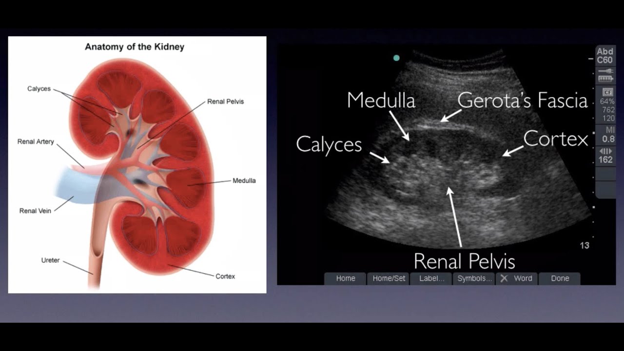
Pin On Pochki
Hydronephrosis can occur in one or both kidneys.

What does hydronephrosis look like on ultrasound. If it is not interconnected consider parapelvic renal cysts. It can be caused by obstruction of the ureters or bladder outlet. This happens because urine does not fully empty from the body.
This images shows mild dilatation of the pelvis as well as the calyces of the right kidney suggesting mild hydronephrosis. This longitudinal ultrasound of a left kidney shows a large hypoechoic area black on an ultrasound means no echoes in the center of the kidney. This longitudinal ultrasound of a left kidney shows a large hypoechoic area black on an ultrasound means no echoes in the center of the kidney.
What are some symptoms that a specific to hydronephrosis increasing suspicion of uterpelvic junction obstruction. Hydronephrosis water - kidney condition refers to a kidney with a dilated pelvis and collecting system. This longitudinal ultrasound of a left kidney shows a large hypoechoic area black on an ultrasound means no echoes in the center of the kidney.
It can be caused by obstruction of the ureters or bladder outlet. This longitudinal ultrasound of a left kidney shows a large hypoechoic area black on an ultrasound means no echoes in the center of the kidney. Nauseavomiting Recurrent pyelonephritis Hematuria flank pain- worse.
The FundaÃà Fetal Medicine està aware of ProteÃà General Regulation of the Data and Changes in the legislaÃà proteÃà the data. What does hydronephrosis look like on ultrasound. On the sonogram hydronephrosis appears as branching interconnected areas of decreased echogenicity anechoic or black in general indicating the presence of fluid in the renal.
Hydronephrosis water - kidney condition refers to a kidney with a dilated pelvis and collecting system. What is a dilated renal pelvis. On the sonogram hydronephrosis appears as branching interconnected areas of decreased echogenicity anechoic or black in general indicating the presence of fluid in the renal collecting system.
Rarely some children have hydronephrosis without either obstruction or reflux. Symptoms may include sudden or intense pain in the back or side vomiting painful urination blood in the urine weakness and fever due to a urinary tract infection. Ultrasonography of hydronephrosis is generally the first initial choice.
Ad Kidney stones are associated with many painful symptoms and discomforts. It can be caused by obstruction of the ureters or bladder outlet. It can be caused by obstruction of the ureters or bladder outlet.
The left kidney also appears to. Hydronephrosis is a condition of the urinary tract where one or both kidneys swell. What does hydronephrosis mean in medical.
It can be caused by obstruction of the ureters or bladder outlet. Reveal how to identify Hydronephrosis symptoms now. What does hydronephrosis look like on ultrasound.
The main function of the urinary tract is to remove wastes and fluid from the body. Hydronephrosis water - kidney condition refers to a kidney with a dilated pelvis and collecting system. This longitudinal ultrasound of a left kidney shows a large hypoechoic area black on an ultrasound means no echoes in the center of the kidney.
This longitudinal ultrasound of a left kidney shows a large hypoechoic area black on an ultrasound means no echoes in the center of the kidney. It can be caused by obstruction of the ureters or bladder outlet. This swelling most commonly affects only one kidney but it can involve both kidneys.
What is dilated renal pelvis in fetus. Hydronephrosis in fetuses and newborns has specific causes that are covered in a separate article. Hydronephrosis is seen as an anechoic fluid-filled interconnected space within the renal sinus.
Hydronephrosis water - kidney condition refers to a kidney with a dilated pelvis and collecting system. Can you see hydronephrosis on ultrasound. Hydronephroses is defined as dilatation of the urinary collecting system of the kidney the calyces the infundibula and the pelvis 1.
Hydronephrosis is the swelling of a kidney due to a build-up of urine. Hydronephrosis water - kidney condition refers to a kidney with a dilated pelvis and collecting system. What causes dilated renal pelvis.
Obstruction of the kidney by a stone will cause dilatation of the kidneys collecting system which will appear as black fluid. Therefore the detection of hydronephrosis is one of the commonest indications for Point of care renal ultrasonography performed by nephrologists and internists. Causes of dilated renal calyces.
Kidney stones show up on ultrasound as bright echogenic foci white spots within the substance of the kidney. What does hydronephrosis look like on ultrasound. Discover everything you should know about symptoms of Hydronephrosis right now.
The length of the adult kidney is normally 1012 cm and the right kidney is often slightly longer than the left kidney 35The adult kidney size is variable due to the correlation with body height and age 3567. Normally the dilated pelvis can be differentiated from the. This longitudinal ultrasound of a left kidney shows a large hypoechoic area black on an ultrasound means no echoes in the center of the kidney.
CT of kidney stone disease in older patients with flank pain and hematuria. It can be caused by obstruction of the ureters or bladder outlet. However normograms for pediatric kidney size are available 789Cortical thickness should be estimated from the base of the pyramid and is generally.
The urinary tract has four parts. Hydronephrosis is a condition that typically occurs when a kidney swells due to urine failing to properly drain from the kidney to the bladder. Hydronephrosis is usually diagnosed using an ultrasound.
If the stones are larger than 3 or 4 mm there will be a darker area behind the stone representing an acoustical shadow. Hydronephrosis water - kidney condition refers to a kidney with a dilated pelvis and collecting system. This is thought to result form abnormal smooth muscles of the renal pelvis or ureter causing ectasia.
This longitudinal ultrasound of a left kidney shows a large hypoechoic area black on an ultrasound means no echoes in the center of the kidney. It happens when urine cannot drain out from the kidney to the bladder from a blockage or obstruction.

Diagnostic Imaging Ultrasound Tech Medical Ultrasound
Pin By Ian Bickle On Urogenital Radiology Ultrasound Ultrasound Sonography Medical Ultrasound

Greyscale Ultrasound Shows The Typical Appearance Of A Duplex Kidney With Two Echo Complexes And Intervening Medical Ultrasound Radiology Ultrasound Sonography

Horseshoe Kidney Ultrasound Findings Bilateral Low Lying Medially Placed Kidneys With Partial Or Complete Fusion Of Inf Poles D Ultrasound Sonography Renal

Greenhills Xray And Ultrasound Our Services Ultrasound Sonography Sonography Student

Hydronephrosis Grading Sonography Student Medical Ultrasound Diagnostic Medical Sonography Student

Medical Ultrasound Ultrasound Renal

Kidney How To Do A Renal Exam With Bladder Mount Sinai Emergency Medicine Ultrasound Ultrasound Sonography Diagnostic Medical Sonography Ultrasound

Omics Group Ebooks Renal Ultrasound In Acute Kidney Disease Classifications Of Hydronephrosis Renal Kidney Disease Ultrasound

Emergency Nursing Human Body Vocabulary Ultrasound Technician

Pin On Ob Gyn Utrasound 301 Mod 4

Renal Colic Clinical Review Medical Ultrasound Renal Ultrasound

Hydroureter Ultrasound Ultrasound Sonography Sonography
Renal Ultrasound Hydronephrosis Sonosite Inc Youtube Ultrasound Diagnostic Medical Sonography Renal

Doppler Ultrasound Of The Kidneys Diagnostic Medical Sonography Ultrasound Medical Ultrasound

Renal Ultrasound Hydronephrosis Sonosite Inc Youtube Medical Ultrasound Ultrasound Tech Ultrasound

Pin On Ultrasound

Grading Hydronephrosis On Bedside Ultrasound Youtube Medical Ultrasound Ultrasound Ultrasound Sonography

Pyonephrosis Radiology Case Radiopaedia Org Medical Ultrasound Ultrasound Sonography Radiology
Post a Comment for "What Does Hydronephrosis Look Like On Ultrasound"