Baby Heart During Ultrasound
The nurse called back the same day and said the doctor said everything looked good normal ultrasound. Hearing a developing babys heartbeat is one of the milestones of pregnancy.

Ultrasound Evaluation Of The Fetal Heart Radiology Key Obstetric Ultrasound Ultrasound Radiology
Or you can listen to it during your first Doppler ultrasound which is a part of your first prenatal check-up scheduled between the 12th and 14th week or later in some cases 3.

Baby heart during ultrasound. The TV host shared a video Tuesday on Instagram of the couple listening to the babys heartbeat during an at-home ultrasound. Heart problems are the most common congenital birth defects and its easy to panic when something unusual is detected on ultrasound. It is a common finding in normal babies.
We know typically your babys heart-beatrate is first visible on sonography around 6 weeks of gestation and we know you cant wait to see and hear the awesome growth occurring inside your body. Normal Fetal Heart Ultrasound. It is not a heart defect and does appear to increase the babys risk of having a heart defect.
Confirm viable pregnancy and check for non-viable molar or ectopic pregnancy confirm babys heartbeat measure babys crown-to-rump length which can help determine gestational age assess. They looked at everything with the baby. It is NOT an abnormality and is NOT associated with heart defects.
Down syndrome can be detected early on in pregnancy with a blood screening of the mother. But echogenic intracardiac focus EIF is almost never something to worry about. Had my first ultrasound last week at 5 weeks 4 days and saw a gestational and yolk sac with small fetal pole.
You can even see and measure this flicker of light on an ultrasound. To do this the ultrasound probe transducer is fastened to your belly. The first thing you need to know is that this finding is usually not an indication of abnormal cardiac anatomy and usually is a normal finding.
Transducer selection should be guided by the acoustic characteristics of. These guidelines have been endorsed by the New Zealand Fetal Maternal Medicine Network NZMFMN and the New Zealand Branch of the Australasian Society for Ultrasound in Medicine ASUM. It shows up as a bright spot on the heart in imaging and its thought to be a microcalcification on the heart muscle.
My second ultrasound is scheduled for Thursday to check for heartbeat and Im terrified. So I went in today. We give God all GLORY for this great.
And hearing it at every prenatal visit is comforting. Dec 21 2021 at 538 PM. On our six week ultrasound we heard our sweet babys heartbeat.
Calcium deposit on heart in ultrasound My daughter is 22 weeks pregnant and the ultrasound showed a white spot on babys heart Calcium deposit on babys liver Down Syndrome White Spots on Heart echogenic focus in the heart. This bright spot is usually first detected during the second-trimester anatomy ultrasound and many parents are naturally concerned about what it means. In the past parents worried that fetal heart rate or a white spot echogenic intracardiac foci on an ultrasound may be related to Down syndrome but these factors are not necessarily associated with a Down syndrome diagnosis.
Possible down syndrome pregnancy for my daughter. Could it be Down syndrom Dr found a spot on my sons heart in ultrasound. It may also be used to check the fetal heart rate during labor.
The continuity of this vascular connection during fetal life is the ductal arch which is flat uniform and slightly larger than the aorta. It sends the sounds of your babys heart to a computer. The presence of an EIF does vary with ethnicity and can be seen in up to 30 of babies of Asian ancestry.
So I had a check up yesterday Im 18 weeks and I had an ultrasound last week because I was spotting a little. Approximately 5 of all normal babies will have an EIF detected during a routine mid trimester ultrasound. The approximate components of the sagittal view of the ductal arch are.
Attaches to the heart valve. During your first ultrasound appointment the doctor or ultrasound technician will check for the following. An irregular fetal heartbeat typically does not indicate long-term issues.
The doctor uses a Doppler ultrasound to gauge the fetal heart rate the number of times the babys heart beats per minute and makes sure it falls within the. We are so thankful to have a strong heartbeat of 155bpm. But it may very mildly increase the risk that the fetus has Down Syndrome.
The truth is your babys heart will likely start beating sometime around week 6 of your pregnancy. This is the farthest weve gotten after 3 years of IVF and 6 failed transfers. Right ventricle main pulmonary artery ductus arteriosus and descending aorta.
In and of itself it will not cause the baby any problems. The healthcare provider may also check your babys heart rate continuously during labor and birth. This document summarizes the recommended techniques for fetal heart assessment.
If you go for an early ultrasound scan around the 6th week you will get to hear as well as see the babys heart beating 2. If you are young healthy and with no family history of DS your chances of a sick baby are 1 in 360000 if an ultrasound spots a deposit on your babies heart the chances of it being DS are 1 in 300000 so even thought your chances are bigger they are still very rare. On ultrasound it looks like a bright spot in the muscle of one of the heart valves.
White spot on babys heart during ultrasound. You wait until you think you are 6 weeks and you call Pregnancy Treasures so we can let you hear that awesome heartbeat via our ultrasound.

The Fetal Doppler Used To Listen To The Baby Heart Beat In Prenatal Visits Dr Gulbahar Donn Md Obgyn Queens Ny Fetal Doppler Prenatal Visits Fetal

Labelled Fetal Heart Ultrasound Ultrasound Fetal Ultrasound Sonography

A Gallery Of High Resolution Ultrasound Color Doppler 3d Images Fetal Heart Ultrasound Fetal Color

The Fetal Three Vessel And Tracheal View Revisited Semantic Scholar Obstetric Ultrasound Cardiac Sonography Ultrasound

Color Doppler Sonography In Assessment Of The Fetal Heart Sonography Fetal Ultrasound School

Cardiac Chambers The Four Chamber And Short Axis Views Obgyn Key Obstetric Ultrasound Medical Ultrasound Ultrasound Sonography

Images Ultrasound Cardiac Sonography Diagnostic Medical Sonography

Pin By Alexandra Ashley On Ultrasound Ultrasound Cardiac Sonography Obstetric Ultrasound

Fetal Heart Ultrasound How To Diagnostic Medical Sonography Ultrasound Obstetric Ultrasound
Obstetric Ultrasound Ultrasound Ultrasound Technician

The Basic Fetal Heart Scan Fetal Scan Basic

4 Chamber View Basal Ultrasound Fetal Color

Fetal Echocardiography Ventricular Septal Defects Youtube Greggory Devore Ventricular Septal Defect Fetal Sonography

Pin On Pregnant

Ultrasound Evaluation Of The Fetal Heart Radiology Key Ultrasound Medical Ultrasound Diagnostic Medical Sonography
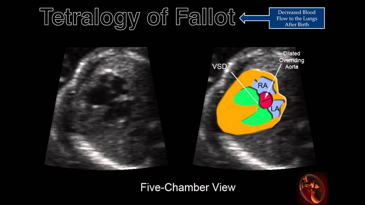
Fetal Echocardiography Tetralogy Of Fallot Tetralogy Fetal Heart Structure

Image Result For Right Outflow Tract Ultrasound Ultrasound Technician Medical Ultrasound Obstetric Ultrasound

Cardiac Chambers The Four Chamber And Short Axis Views Obgyn Key Medical Ultrasound Cardiac Sonography Obstetric Ultrasound

Pin By Luca Szadovszky On Cytogenetic Ultrasound Fetal High Resolution
Post a Comment for "Baby Heart During Ultrasound"