Where Is The Baby In An Ultrasound
This may be harder to see with some babies which is why there are multiple signs to look for during an ultrasound. Increasingly cleft lip and cleft palate are seen on ultrasound before the baby is born.

Peace On Earth Funny Babies Ultrasound Funny Pictures
An ultrasound is a type of technology that uses sound waves to create images.

Where is the baby in an ultrasound. By 8 weeks gestation your baby can usually be seen with transabdominal ultrasoundBy 10-11 weeks gestation the embryo is clearly recognisable as a baby with a body head arms and legs as well as many other identifiable features. Ultrasound examinations provide parents with a valuable opportunity to view and hear the heartbeat of the fetus bond with the unborn baby and capture images to share with family and friends. This is where you can see the tip of the penis peeking out from behind the testicles.
A pregnancy ultrasound is an imaging test that uses high frequency sound waves to create pictures of a baby in the womb as well as the mothers READ MORE 5 Weeks Pregnant. Your baby may be moving around the pregnancy sac. Its the first thing to develop inside the gestational sac.
A Meet the Baby Ultrasound is safe for both mother and fetus. But a fetus growing inside a liver is shocking even to medical professionals. As for the 4 week ultrasound there are a few things the ultrasound can and cannot show.
Your baby will also look grey or whitish and will be located within the amniotic fluid the dark area inside of the womb. The yolk sac functions as a means for the nourishment of the embryo before the circulatory system and the placenta develop. It creates an image of the baby in the mothers womb uterus.
A mid-pregnancy anatomy ultrasound takes longer about 20 to 45 minutes because the sonographer will look closely at your babys basic anatomy including the head brain face neck chest heart spine stomach kidneys bladder arms legs and umbilical cord so your provider can make sure theyre developing properly. The diagnosis is then confirmed at birth with a detailed visual assessment and physical examination. Its a safe way to check the health of an unborn baby.
In most cases a prenatal ultrasound can detect cleft lip alone or cleft lip and palate as early as 16 weeks into a pregnancy. The details that you see in the image will depend on the stage of your pregnancy. Your ultrasound may only show a small circle at the center which is called as a gestational sacAt this stage it is not possible to see much more than the gestational sac and the sac is identifiable until you are 4 ½ weeks pregnant.
Our state of the art technology uses the EXACT same ultrasound waves used in a traditional 2D ultrasound scan at your doctors office. Even before you can see an embryo inside the gestational sac you should spot the yolk sac. A TikTok from Dr.
Spot the baby. The machine and software simply process the information differently to provide us with spectacular 3D ultrasound images and 4DHDlive video. Ultrasound at 2 months pregnant In a healthy pregnancy you can expect to see some cool things during a transvaginal ultrasound.
Measurements of the yolk sacs size and shape are important when assessing the pregnancy. By conducting an ultrasound scan in the sixth week of pregnancy the doctor is able to tell about the location of your baby. Your baby is also going to look either grey or white on the ultrasound image.
During a fetal ultrasound the babys heart head and spine are evaluated along with other parts of the baby. Look at the area within your amniotic fluid to try to make out the outline and features of your baby. It is during this scan she will be able to determine whether the baby is in an ideal location or not.
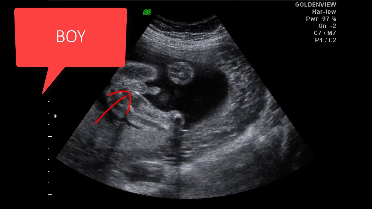
2 days agoSome people may have heard of an ectopic pregnancy where a fetus grows outside the womb. It can be seen on ultrasound between the embryo and the gestational sac. When the ultrasound technician is looking for a boy they are looking for something called the turtle sign.
Fetal ultrasound is a test used during pregnancy. The gestational sac the yolk sac the early shape of your baby and their super-fast heartbeat. Baby has long legs in ultrasound Arms and legsBetween 5 and 6 weeks your baby sprouts two flipper-like buds that will lengthen and grow into arms and by 7 weeks two additional buds form that will become his legsYour babys hands and feet which look somewhat like paddles at this stage will form at the end of these buds.
It emits sound waves that bounce off of your babys tissues fluids and bones. When is cleft palate diagnosed. During pregnancy a transducer or wand is placed in your vagina or on top of your belly.
Baby will be situated in the amniotic fluid which as we have already discussed will be the dark area on the image. The details that you will see when reading the ultrasound will vary greatly depending on the stage of your babys development and your pregnancy. It is not possible to measure fetal size at this stage.
Regarding this when can an abdominal ultrasound be done in pregnancy.

Pin On Pregnant

18 Week Obgyn Visit And Ultrasound Girl Ultrasound Pictures Baby Ultrasound Pictures Ultrasound Pictures

20 Week Ultrasound Why Is It Important Baby Ultrasound Pictures Ultrasound Pictures 20 Weeks Pregnant Ultrasound

Pin On Baby By Weeks

Baby S First Ultrasound 11 Weeks 1 Day First Ultrasound Girl Ultrasound Pictures Boy Ultrasound Pictures

Baby Gender Using Nub Theory Baby Gender Prediction Baby Ultrasound Baby Gender
Pin On Gender

The 12 Week Scan When You See Your Real Actual Child For The First Time Is The Most Awe Inspiring Thing You Ll Witness Girl Ultrasound Pictures Ultrasound Pictures 12 Week Scan

When Will I Get My First Ultrasound Pictures Trimester Talk First Ultrasound Baby Ultrasound Ultrasound Pictures

14 Weeks Pregnant 2d Ultrasound Active Baby 14 Weeks Pregnant 14 Weeks Pregnant Ultrasound 15 Weeks Pregnant

My Brother S Best Friend Chapter 4 In 2021 Girl Ultrasound Pictures Ultrasound Pictures Baby Ultrasound Pictures

Pin On Pregnant

Jessa Duggar Shares Ultrasound Picture Of Baby Number 3 Duggars Baby Pictures Baby Number 3

Pin On Fetal Sex

15 Weeks 5 Days Ultrasound Ultrasound Baby Boy

Baby Ultrasound Pictures Baby Ultrasound Baby Ultrasound Pictures Ultrasound Pictures

With Today S Advanced Technology We Have A Real Time Window Into Development And Play In The Womb Baby Ultrasound Boy Ultrasound Pictures Ultrasound Pictures

5 Month Old Or 23 Weeks Baby Ultrasound Baby Ultrasound Baby Month By Month 5 Month Old Baby
Pin On Baby Development Week By Week
Post a Comment for "Where Is The Baby In An Ultrasound"