How To Understand Baby Ultrasound
An ultrasound relies on making a visual representation of what is going on inside of your womb. If youre 12 weeks along in the pregnancy you may be able to make out your babys head and if youre 20 weeks along you may even see the spine heart feet and eyes.

Pin On Pregnant
Some tissues absorb sound waves while others reflect them.

How to understand baby ultrasound. Ad Learn these shocking facts safety and benefits of ultrasound imaging now. So the first step to help you read the ultrasound image is to be familiar with the anatomy that you are imaging. 1 However there are certain signs in boy.
For a clearer ultrasound measurement there are newer technologies that offer more detailed images. It is necessary to understand that the width of the image is equal to the probe footprint size only at the uppermost part of the image and the depth marks on the side of the screen are pertinent only for measurement of the depth on the line drawn through the middle of the probe. Also note that the solider tissue will look whiter on.
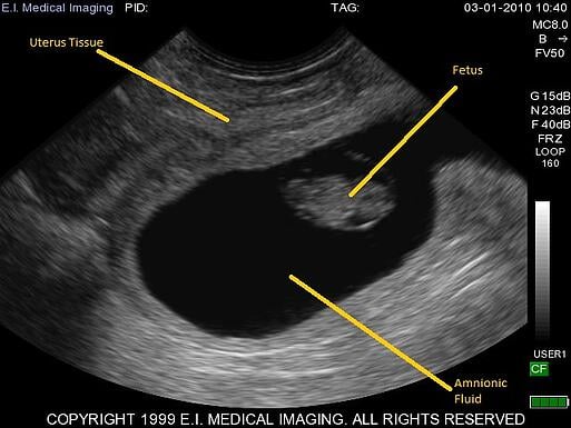
You need to understand that white is solid and black is liquid. Fetal ultrasound measurements can show how the baby is growing and detect abnormalities. An ultrasound or sonogram picture is a black and white photograph so they all look the same to someone who doesnt know much about how to read an ultrasound.
The accuracy of the report will depend on many factors including the age of the baby the equipment used the technician and the baby. This understanding will make it easier for you to see other details in an ultrasound image. An ultrasound machine uses sound waves for scanning the abdominal area of a woman.
During pregnancy many different ultrasounds measurements can be done. Find out which illnesses and disease can be caught from an ultrasound today. A baby sonogram also known as an ultrasound is a common procedure during a pregnancy.
Fetal ultrasound measurements can include the crown-rump length CRL biparietal diameter BPD femur length FL head circumference HC occipitofrontal diameter OFD abdominal. Understanding how an ultrasound can be used to tell your babys sex is an important part of knowing how reliable the ultrasound will be. The most common type of ultrasound is a 2-D ultrasound which produces a two-dimensional image of your baby.
Ultrasounds guide future mums during the whole pregnancy process and are essential to understand how the baby progresses thanks to the ultrasound pregnancy is confirmed and is possible to know the number of babies they are expecting ruling out possible fetal abnormalities and reveal the fetal growth. To read an ultrasound picture look for white spots on the image to see solid tissues like bones and dark spots on the image to see fluid-filled tissues like the amniotic fluid in the uterus. Sonograms emit high frequency sound waves from a transducer placed on the mothers abdomen.
Typically the ultrasound is done halfway through the pregnancy. It is called the fetal anatomy survey and it is done to look for fetal anomalies not simply to find out the sex of your baby. The density of the tissue dictates the speed at which the echoes return.
The waves detect the foetus and produce a scanned image called sonogram. By measuring the sonogram the technician can predict the age and the development of the baby almost accurately. There is also a 3-D ultrasound that allows doctors to see the width height and depth of the fetus and your organs Healthline states that this type of ultrasound can help a doctor see and diagnose any suspected problems throughout your.
Read an ultrasound picture an ultrasound may be performed for a variety of reasons but looking at a baby in the womb is the most common reason. The fetus is scanned by rays of repetitive ultrasound beams that reflect back to the transducer and onto a monitor screen. Various body tissues conduct sound differently.
Using an ultrasound you can usually see your babys heartbeat at around 8 weeks of pregnancy for example. Youtubersakhi subsribemy other vdoshow to read baby ultrasound anomaly2 d levelhttpsyoutubewcMOu_1OsX4बन अलटरसउड. Robyn Horsager-Boehrer explains step-by-step what obstetricians are looking for when they conduct 18- to 20-week ultrasounds on pregnant women.

Baby Gender Using Nub Theory Baby Gender Prediction Baby Ultrasound Baby Gender

Pin On Pregnancy

Pin On Ultrasound

Pin On Precho Babyechografie Antwerpen

Not Found First Ultrasound Baby Gender Ultrasound Boy Ultrasound Pictures

6 Ways To Tell Baby S Gender From An Early Sonogram Cafemom Com

20 Weeks Old Jo Mcvey Photography Nashville Child And Family Photographer Future Baby New Baby Products Baby Pictures

Understanding Your Fetal Ultrasound Youtube

Pin On Diary Of A Fit Mommy Sia Cooper

The Dangle Angle Girl Or Boy Ultrasound Gender Pregnant Gender Prediction

How To Read An Ultrasound Picture Go Life Mobile Medical Inc

How To Read An Ultrasound Picture 9 Steps With Pictures

Pin On Pregnancy

Pin On Radiology

See Your Babys Features In 2d 3d And Hd Live Motion And Understand The Difference At Miracle Inside We Offer 2d3d Baby Scan 4d Baby Scan Baby Ultrasound

Pin On Baby Time
Ultrasound Basics How To Read An Ultrasound Image
Pin On Ultrasoundfeminsider Com

Pin On Our New Bundle Of Joy
Post a Comment for "How To Understand Baby Ultrasound"