Ultrasound Baby Heart Defect
We are able to detect the majority of major heart defects with ultrasound Common heart abnormalities explained The most common congenital heart abnormality is a ventricular septal defect VSD says Dr. This type of VSD is called a ventricular septal defect.
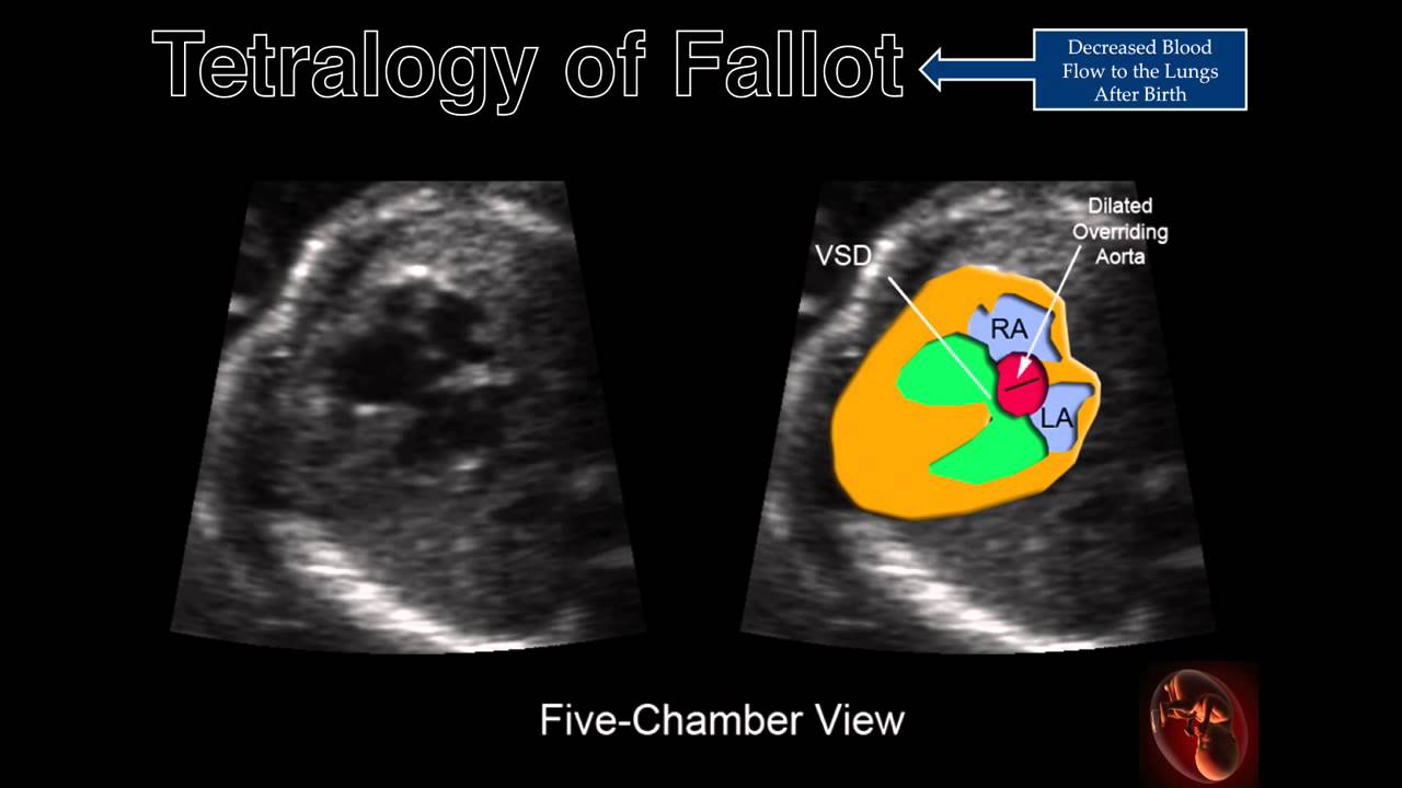
Fetal Echocardiography Tetralogy Of Fallot Tetralogy Fetal Heart Structure
It might be nothing to worry about but you definitely want to get it checked.

Ultrasound baby heart defect. The news comes as a shock. This tool shows movement over time which is displayed on the bottom part of the image. When can a heart defect be detected in a baby with Down syndrome.
Diagnosis of fetal heart defects in particular can improve newborn outcomes and enable further research on in utero therapies the researchers said. If a sonographic exam suggests the defect a secondary amniocentesis can usually confirm the chromosomal anomaly with a high degree of accuracy. Detection of fetal VSD is done by cardiac ultrasound performed between 18 and 22 weeks of pregnancy.
In some cases an ultrasound may raise concerns about a problem but not offer enough information to make a definitive diagnosis. If a first degree relative has been diagnosed with a congenital heart defect. A fetal echo may be performed by the maternal fetal.
Fetal cardiac examination is an indispensable part of the prenatal ultrasound because of the following well-recognized reasons. The sound waves can also detect blood flow throughout the babys heart. First degree relative includes the mother or father of the baby as well.
Do ultrasound pictures show heart rate. When we carried out a prenatal ultrasound scan in the Fetal Cardiology clinic we confirmed that the structure of your babys heart is normal but there is a small hole between the two ventricles. These ultrasound and color Doppler images show a VSD in the muscular part of the ventricular septum of the fetal heart.
Fetal echocardiograms are recommended for the following women. 1 The incidence increases to 751000 live births if all mild. To evaluate prospectively the efficacy to screen for congenital heart defects CHD during the first trimester nuchal translucency NT ultrasound examination by assessing the four chambers view of fetal heart.
Babies do breathe while in utero but once they are born there is a chemical released that closes a particular valve in the heart that is no longer needed. Ad Discover these surprising facts about ultrasound imaging right now. Sonography of fetal VSD.
Should a prenatal ultrasound indicate your baby may have a heart defect or if you have risk factors your obstetrician will most likely order a test called a fetal echocardiogram to examine your babys heart before birth. The image on the bottom shows how the babys heart rate is calculated. One such example is when Down syndrome is suspected.
First congenital heart diseases CHDs are common congenital anomalies. Flow is seen across the defect in the septum from left ventricle to the right color Doppler images. This enables the doctor to evaluate the structure and function of the fetal heart.
Pregnancies that were examined prospectively by ultrasound in the first trimester 11th-14th week the second 19th-24th week and third. Down syndrome can be detected early on in pregnancy with a blood screening of the mother. A fetal echocardiogram echo is a detailed ultrasound exam that takes images of the babys heart.
The incidence of moderate to severe forms of CHD is about 61000 live births. We refer to this as a ventricular septal defect VSD the word septal refers to the wall between the two sides of the heart. Fetal ultrasound screening is universally recommended during the second trimester of pregnancy in the United States and by the World Health Organization.
Sound waves ultrasound are used in this test to produce a moving image of the heart. Many defects are discovered when an ultrasound picks up a fetal heart condition. The size of the heart more than doubles from 13 weeks to 19 weeks therefore there are anomalies which may not be diagnosed at the early structural scan.
The top part of the image shows placement of a measuring tool on the ultrasound machine called an M-mode through the image of the beating heart. During your routine 20-week pregnancy ultrasound your physician notices a potential abnormality with your babys heart. Doppler 2D USG for congenital heart defects.
In the past parents worried that fetal heart rate or a white spot echogenic intracardiac foci on an ultrasound may be related to Down syndrome but these factors are not necessarily associated with a Down syndrome. The heart is a rounded structure and should be assessed from multiple angles and positions to ensure that even the smallest abnormality is not missed. Due to her defect she actually needed that value to stay open so she needed to get medication immediately to keep the valve from closing.

Cardiac Chambers The Four Chamber And Short Axis Views Obgyn Key Medical Ultrasound Diagnostic Medical Sonography Obstetric Ultrasound

Tetralogy Of Fallot Pulmonary Atresia With Ventricular Septal Defect And Absent Pulmonary Valve Sy Ventricular Septal Defect Ultrasound Sonography Ultrasound

Ultrasound Evaluation Of The Fetal Heart Radiology Key Ultrasound Ultrasound Sonography Obstetric Ultrasound

Atrial Ventricular And Atrioventricular Septal Defects Obgyn Key Diagnostic Medical Sonography Ultrasound Ventricular Septal Defect

Fig 2 Pathological Specimen Of The V Shaped Great Vessels With The Aortic Arch Ao Junction With Diagnostic Medical Sonography Ultrasound Cardiac Sonography

6 Questions Congenital Heart Defect Awareness Chd Awareness Congenital Heart Defect
Ultrasound Obstetric Ultrasound Diagnostic Medical Sonography

Cardiac Chambers The Four Chamber And Short Axis Views Obgyn Key Obstetric Ultrasound Medical Ultrasound Ultrasound Sonography

A Gallery Of High Resolution Ultrasound Color Doppler 3d Images Fetal Heart Ultrasound Fetal Color

Fetal Echocardiography Ventricular Septal Defects Youtube Greggory Devore Ventricular Septal Defect Fetal Sonography

Cardiac Chambers The Four Chamber And Short Axis Views Obgyn Key Ultrasound Cardiac Sonography Diagnostic Medical Sonography

Hypo Plastic Left Heart Syndrome Medical Ultrasound Diagnostic Medical Sonography Ultrasound Technician

Pin By Roslyn Ramirez On Ecografias Ultrasound Technician Ultrasound Obstetric Ultrasound

Ultrasound Of Atrial Septal Defects Ultrasound Diagnostic Medical Sonography Obstetric Ultrasound

Cardiovascular System Diagnosis Of Congenital Abnormalities The 18 23 Weeks Scan Cardiovascular System Fetal Diagnosis

Ask The Right Questions At The A S Congenital Heart Congenital Heart Defect Heart Defect

Ultrasound Evaluation Of The Fetal Heart Radiology Key Obstetric Ultrasound Ultrasound Radiology

Fetal Heart Ultrasound How To Diagnostic Medical Sonography Ultrasound Obstetric Ultrasound

Atrial Ventricular And Atrioventricular Septal Defects Obgyn Key Ultrasound Congenital Heart Defect Atrial Septal Defect
Post a Comment for "Ultrasound Baby Heart Defect"